Juvenile Spring Eruption: What You Need To Know
A common sun-induced rash on ears in boys during early spring, self-limiting with proper care and prevention.

Juvenile spring eruption (JSE) is a distinct photodermatosis characterized by itchy, papulovesicular lesions primarily on the ears of boys and young men following early spring sun exposure. It typically resolves within 1-2 weeks without scarring and is considered a localized variant of polymorphous light eruption (PMLE).
What is juvenile spring eruption?
**Juvenile spring eruption** is a self-limiting skin condition triggered by ultraviolet (UV) light exposure, most commonly affecting the light-exposed skin of the ears. It manifests as multiple small, red, itchy papules that evolve into vesicles, blisters, and crusts. The condition predominantly impacts boys and young men aged 4-25 years, likely due to less hair coverage on their ears compared to girls.
Outbreaks occur abruptly 8-24 hours after initial sun exposure in early spring or occasionally early summer, particularly under sunny but cold weather conditions. Lesions heal spontaneously in about 2 weeks, often faster with treatment, and recurrences are seasonal without long-term sequelae. Enlarged cervical lymph nodes may accompany the eruption in some cases.
Who gets juvenile spring eruption?
JSE primarily affects
Caucasian boys and young men
in temperate climates, with peak incidence in children aged 7-12 years. Males are more susceptible because their ears are less protected by hair, and protruding ears increase vulnerability. Family history of photosensitivity or PMLE may predispose individuals.- Age: Typically 4-25 years, resolving by puberty or young adulthood in most cases.
- Gender: Overwhelmingly boys (90%+ cases).
- Risk factors: Fair skin, prominent ears, first intense spring sun exposure after winter.
Outbreaks can be epidemic-like in schools during sudden sunny spells in spring.
Clinical features of juvenile spring eruption
The hallmark of JSE is an
acute eruption on the helices and antihelices of the ears
, sparing the earlobes and face. Lesions appear within hours to 1-2 days post-exposure:- Initial phase: Multiple red, oedematous papules (2-5 mm).
- Evolution: Papules become vesicular, bullous, or pustular; may crust or erode.
- Symptoms: Intense pruritus; mild pain or burning.
- Duration: Peaks at 2-7 days, resolves in 7-14 days.
- Associated findings: Posterior cervical lymphadenopathy (20-30% cases); occasional mild fever.
Lesions are monomorphic on ears but resemble PMLE elsewhere if generalized. No systemic symptoms beyond itch.
Pathophysiology of juvenile spring eruption
JSE represents a
delayed-type hypersensitivity reaction
to UV-altered skin antigens, akin to PMLE. UVA and possibly UVB penetrate ear cartilage poorly, localizing damage. Cold weather may exacerbate via vasoconstriction, suggesting perniosis overlap (“spring perniosis”).Histology shows superficial and deep perivascular lymphocytic infiltrate, epidermal spongiosis, and occasional interface changes, distinguishing it from acute PMLE sunburn reaction.
Diagnosis of juvenile spring eruption
Diagnosis is
clinical
, based on characteristic history, seasonal timing, and ear-limited lesions in boys. No routine tests needed. Phototesting rarely indicated.Differential diagnosis
| Condition | Key Distinguishing Features |
|---|---|
| Polymorphous light eruption (PMLE) | Wider sun-exposed areas (face, chest, arms); women > men; recurrent summer. |
| Solar urticaria | Hives within minutes of exposure; resolves <1 hour after shade. |
| Photosensitive eczema | Chronic, persistent; positive photopatch tests. |
| Lupus erythematosus (discoid/cutaneous) | Atrophic scars; positive ANA; persistent. |
| Chilblains (perniosis) | Cold > sun trigger; acrocyanosis; winter. |
| Infectious (impetigo, herpes) | Pustules/vesicles with systemic signs; cultures positive. |
Biopsy if atypical: Confirms lymphocytic vasculitis without thrombosis.
Treatment of juvenile spring eruption
JSE is self-resolving; treatment targets symptoms and speeds healing.
Curative management
- First-line: Potent topical corticosteroids (e.g., mometasone 0.1% ointment) twice daily for 7-10 days. Reduces inflammation and itch.
- Emollients: Barrier creams to protect and hydrate.
- Antihistamines: Oral sedating types (e.g., hydroxyzine) for severe pruritus.
- Severe/recalcitrant: Short-course oral prednisolone (1 mg/kg x 3-5 days).
Avoid triggers during acute phase.
Prevention of recurrences
Essential for seasonal control:
- Sun protection: SPF 50+ broad-spectrum sunscreen on ears daily; water-resistant.
- Physical barriers: Wide-brimmed hats, earmuffs; grow hair over ears.
- Gradual exposure: “Harden” skin with increasing sun time.
- Prophylaxis: Polypodium leucotomos extract (240 mg bid), nicotinamide 500 mg bid pre-season (evidence from PMLE).
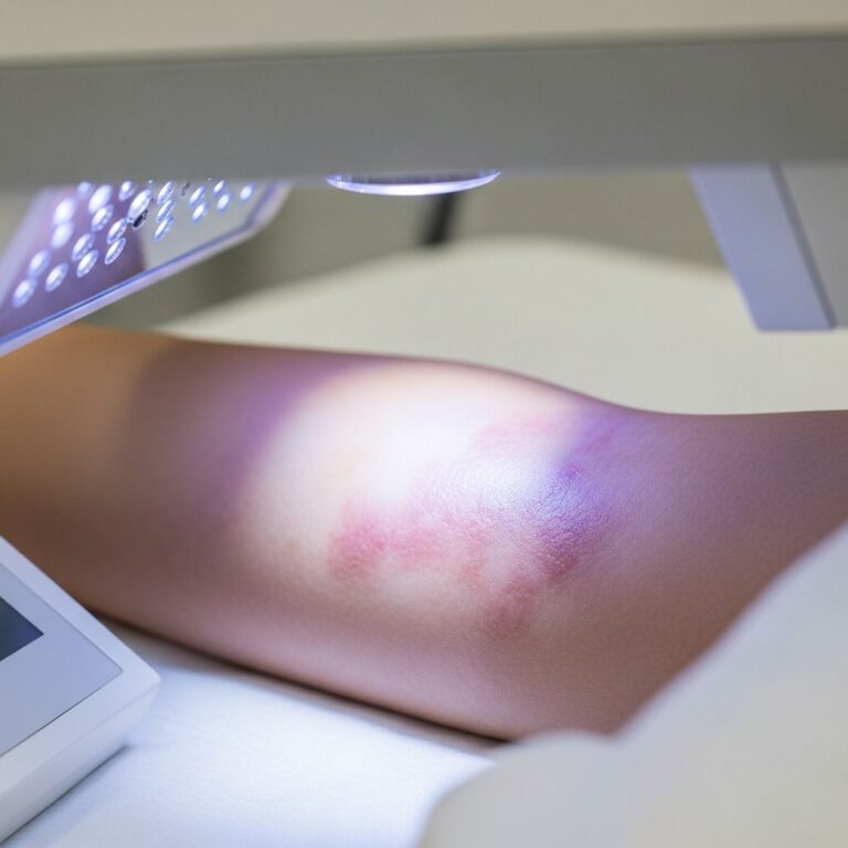
- Phototherapy: Narrowband UVB 2-3x/week x 4-6 weeks pre-spring for frequent cases.
Prognosis and complications of juvenile spring eruption
Excellent prognosis: Lesions heal without scarring in 95% cases. Recurrences decrease with age, resolving post-puberty. Rare milia or post-inflammatory hyperpigmentation. No progression to chronic photosensitivity.
Frequently Asked Questions
Is juvenile spring eruption contagious?
No, JSE is not infectious; it’s an immune reaction to sun exposure.
Does juvenile spring eruption only affect ears?
Primarily yes, but rare cases involve cheeks or neck if PMLE overlap.
Can girls get juvenile spring eruption?
Rarely; <10% cases, usually with short hair or prominent ears.
How long does juvenile spring eruption last?
Untreated: 1-2 weeks; with steroids: 3-7 days.
Will my child outgrow juvenile spring eruption?
Yes, most stop recurring by late teens.
Guidelines
- Diagnose clinically in boys with springtime ear rash post-sun.
- Treat promptly with topical steroids + emollients.
- Educate on prevention: Sunscreen, hats essential.
- Reassure families: Benign, self-limited.
- Refer if persistent, widespread, or atypical.
References
- Juvenile spring eruption in a young boy — Contemporary Pediatrics. 2023-04-01. https://www.contemporarypediatrics.com/view/juvenile-spring-eruption-in-a-young-boy
- Juvenile spring eruption — DermNet NZ. Accessed 2026. https://dermnetnz.org/topics/juvenile-spring-eruption
- Juvenile Spring Eruption: A Variant of Perniosis? — American Journal of Dermatopathology. 2015-09. https://pubmed.ncbi.nlm.nih.gov/26291421/
- Polymorphic light eruption & juvenile spring eruption — Primary Care Dermatology Society. 2022-05-28. https://www.pcds.org.uk/clinical-guidance/polymorphic-light-eruption-and-juvenile-spring-eruption
- Juvenile Spring Eruption Associated With Parvovirus B19 Infection — JAMA Dermatology. Accessed 2026. https://jamanetwork.com/journals/jamadermatology/fullarticle/2702470
- Juvenile spring eruption: an outbreak report and systematic review — British Journal of Dermatology. 2013. https://academic.oup.com/bjd/article/168/5/1066/6614353
Similar Articles


Read full bio of medha deb