Kaposi Sarcoma: 4 Types, Symptoms, Diagnosis, Treatment
Kaposi sarcoma: Rare cancer linked to HHV-8, affecting skin and organs, with types from classic to AIDS-related.

Kaposi sarcoma (KS) is a multicentric angioproliferative spindle cell tumour derived from endothelial cells that line blood and lymph vessels. It is one of the more common tumours induced by viruses in humans and probably arises as a result of the virus-induced release of inflammatory cytokines and growth factors that cause proliferation of endothelial cells.
What is Kaposi sarcoma?
Kaposi sarcoma was first described by the Hungarian dermatologist Moritz Kaposi in 1872. It is caused by infection with human herpesvirus 8 (HHV-8), also called Kaposi sarcoma–associated herpesvirus (KSHV). HHV-8 infection is necessary but not sufficient to cause KS; additional factors such as immunosuppression are required for tumour development. The virus is transmitted through saliva, sexual contact, and blood, with higher prevalence in certain populations like those of Mediterranean or African descent.
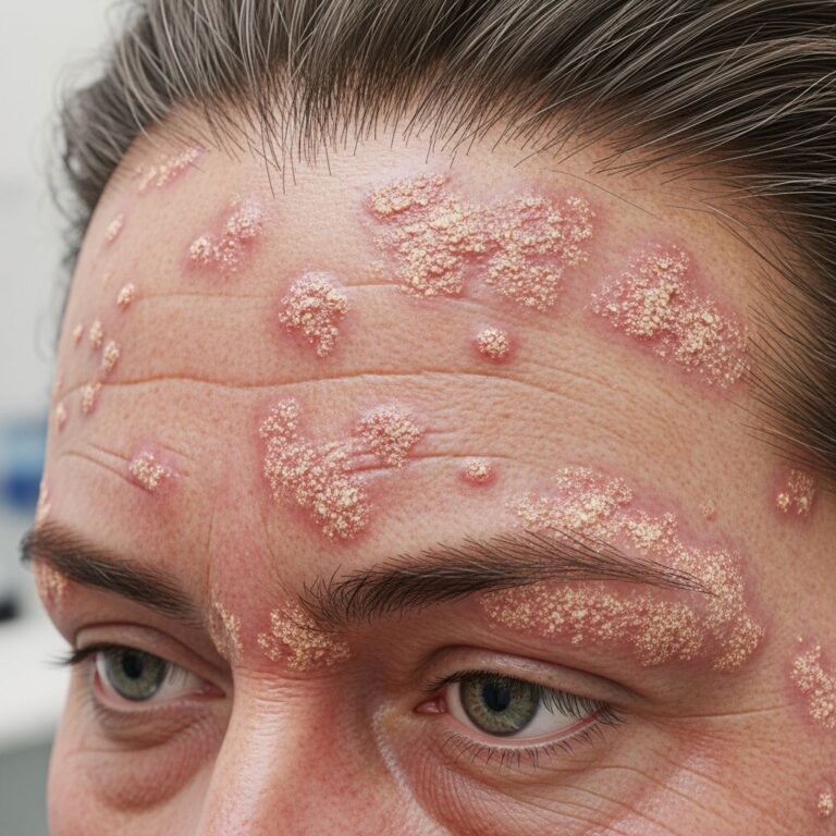
KS manifests as reddish-purple vascular nodules or plaques on the skin, mucous membranes, and internal organs. Lesions can range from small macules to large tumours and may ulcerate or cause swelling due to lymphatic obstruction. While historically rare, KS surged during the HIV/AIDS epidemic, becoming an AIDS-defining illness.
Who gets Kaposi sarcoma?
KS affects specific populations based on its epidemiological subtypes. Risk factors include HHV-8 seropositivity, immunosuppression (e.g., HIV/AIDS, organ transplantation), and genetic predispositions in endemic areas.
- Classic (sporadic) KS: Primarily elderly men of Mediterranean, Eastern European, or Jewish ancestry, aged 50–70 years. Slow-growing lesions on lower legs; better prognosis with indolent course.
- Endemic (African) KS: Common in sub-Saharan Africa, affecting children and adults. Nodular form in adults; aggressive lymphadenopathic form in children with high mortality.
- Immunosuppression-associated (iatrogenic) KS: Occurs in transplant recipients or those on corticosteroids/immunosuppressants. Lesions regress upon reducing immunosuppression.
- Epidemic (AIDS-associated) KS: Most common in HIV-positive individuals, especially men who have sex with men (MSM). Aggressive, multifocal disease; incidence dropped with HAART but persists in advanced AIDS.
Men are affected 10–20 times more often than women in non-AIDS forms. HHV-8 prevalence varies: 5–20% in general population, up to 100% in endemic regions.
What causes Kaposi sarcoma?
The primary cause is HHV-8 infection, which infects endothelial cells and B-lymphocytes, promoting angiogenesis via viral genes like LANA-1 and cytokines (e.g., IL-6, VEGF). Immunosuppression allows viral reactivation and tumourigenesis. In HIV patients, low CD4 counts (<200 cells/μL) strongly correlate with KS development.
Cofactors include HIV co-infection (synergistic), genetic factors (e.g., IL-6 polymorphisms), and environmental triggers like onchocerciasis in Africa. HHV-8 latency persists lifelong, with lytic replication driving lesion progression.
What are the clinical features of Kaposi sarcoma?
KS lesions evolve through patch, plaque, and nodular stages, appearing as red, purple, or brown macules, plaques, or tumours. Distribution varies by subtype.
- Skin: Lower extremities in classic KS; widespread in AIDS-KS. Lesions may be hyperpigmented, oedematous, or ulcerated.
- Mucosal: Oral cavity common (palate, gums); painful, bleeding nodules.
- Visceral: GI tract (bleeding, obstruction), lungs (dyspnoea, haemoptysis), lymph nodes.
Symptoms include pain, swelling (lymphedema), fever, weight loss in advanced disease. Classic KS causes leg oedema; AIDS-KS often asymptomatic initially but progresses rapidly.
How is Kaposi sarcoma diagnosed?
Diagnosis relies on clinical suspicion and confirmed by biopsy. High-resolution dermatoscopy shows rainbow pattern or glomerular vessels.
- Biopsy: Essential; reveals spindle cells, promontory sign, hyaline globules. HHV-8 LANA-1 immunohistochemistry is diagnostic (nuclear positivity).
- Imaging/Staging: CT chest/abdomen for visceral involvement; endoscopy/colonoscopy for GI; bronchoscopy for lungs.
- Other: CD4 count in HIV; HHV-8 serology (supportive).
Staging considers T (tumour extent), I (immune status), S (systemic symptoms). AIDS Clinical Trials Group (ACTG) system guides prognosis.
What is the treatment for Kaposi sarcoma?
No cure exists, but treatments control disease, achieve remission, and improve quality of life. Approach depends on subtype, extent, symptoms, and immunosuppression.
| Type | Treatment Options |
|---|---|
| Localised | Local excision, cryotherapy, intralesional chemotherapy, radiation |
| Advanced/AIDS-KS | HAART + chemotherapy (liposomal doxorubicin), immunotherapy |
| Iatrogenic | Reduce immunosuppression, sirolimus |
- HAART: First-line for AIDS-KS; restores immunity, regresses lesions in 70–90%.
- Radiation: Effective for symptomatic/palliative lesions (47–99% response); electron beam for widespread skin disease.
- Chemotherapy: Liposomal doxorubicin (every 2–3 weeks), paclitaxel for visceral disease.
- Other: Interferon-alpha, targeted (bevacizumab, pomalidomide), topical retinoids.
Observation for asymptomatic classic KS. Multidisciplinary care essential.
Complications
Local: ulceration, secondary infection, cosmetic disfigurement. Lymphedema from lymphatic obstruction. Visceral: GI bleeding (10–40%), pulmonary infiltration (dyspnoea), organ dysfunction. In AIDS-KS, associated with poor prognosis if untreated.
Prevention
Prevent HHV-8 transmission (safe sex, avoid saliva sharing). HAART for HIV prevents epidemic KS. No vaccine yet; antivirals ineffective.
Prognosis
Excellent for classic KS (indolent, >10 years survival). AIDS-KS: good with HAART (median survival >3 years). Poor in untreated visceral/lymphadenopathic forms. Response rates high with modern therapy.
Frequently Asked Questions
Q: Is Kaposi sarcoma curable?
A: No, but manageable with treatments like HAART and chemotherapy, leading to long-term remission in many cases.
Q: Can Kaposi sarcoma be prevented?
A: By preventing HIV and HHV-8 transmission; HAART reduces risk in HIV patients.
Q: What does Kaposi sarcoma look like?
A: Reddish-purple patches, plaques, or nodules on skin, mouth, or mucosa.
Q: Is biopsy always needed?
A: Yes, for definitive diagnosis via histology and HHV-8 staining.
Q: How effective is HAART for AIDS-KS?
A: Highly effective, regressing lesions in most patients by boosting immunity.
References
- Diagnosis & Treatment of Kaposi Sarcoma (KS) — Memorial Sloan Kettering Cancer Center. 2023. https://www.mskcc.org/cancer-care/types/kaposi-sarcoma/diagnosis-treatment-msk
- Kaposi Sarcoma Treatment – NCI — National Cancer Institute. 2024-01-15. https://www.cancer.gov/types/soft-tissue-sarcoma/patient/kaposi-treatment-pdq
- Kaposi sarcoma – Diagnosis and treatment — Mayo Clinic. 2024. https://www.mayoclinic.org/diseases-conditions/kaposis-sarcoma/diagnosis-treatment/drc-20577331
- Kaposi Sarcoma — Merck Manuals Professional Edition. 2024. https://www.merckmanuals.com/professional/dermatologic-disorders/cancers-of-the-skin/kaposi-sarcoma
- Kaposi Sarcoma | Diagnosis & Disease Information — Cancer Therapy Advisor. 2023. https://www.cancertherapyadvisor.com/ddi/kaposi-sarcoma/
- Kaposi Sarcoma – StatPearls — NCBI Bookshelf. 2023-07-17. https://www.ncbi.nlm.nih.gov/books/NBK534839/
Similar Articles


Read full bio of medha deb