Keratitis: Causes, Symptoms, and Treatment
Understanding keratitis: A comprehensive guide to this serious eye inflammation and corneal infection.

What Is Keratitis?
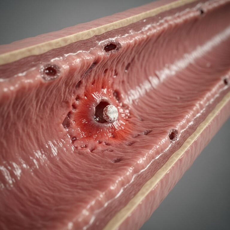
Keratitis is an inflammation of the cornea, the clear front part of the eye that covers the iris and pupil. This condition is considered an ocular emergency and represents one of the major causes of blindness worldwide. The cornea plays a critical role in vision by focusing light onto the retina, and any inflammation or infection affecting it can significantly impact your eyesight. The severity of keratitis ranges from mild surface inflammation to deep corneal infection that can lead to permanent scarring and vision loss if left untreated.
Understanding keratitis is essential because early recognition and prompt treatment can prevent serious complications and preserve vision. The condition can develop rapidly, sometimes progressing from mild symptoms to severe corneal damage within days. This is why any eye pain, redness, or vision changes should be evaluated by an eye care professional immediately.
Types of Keratitis
Keratitis can be classified into several types based on the underlying cause:
Infectious Keratitis
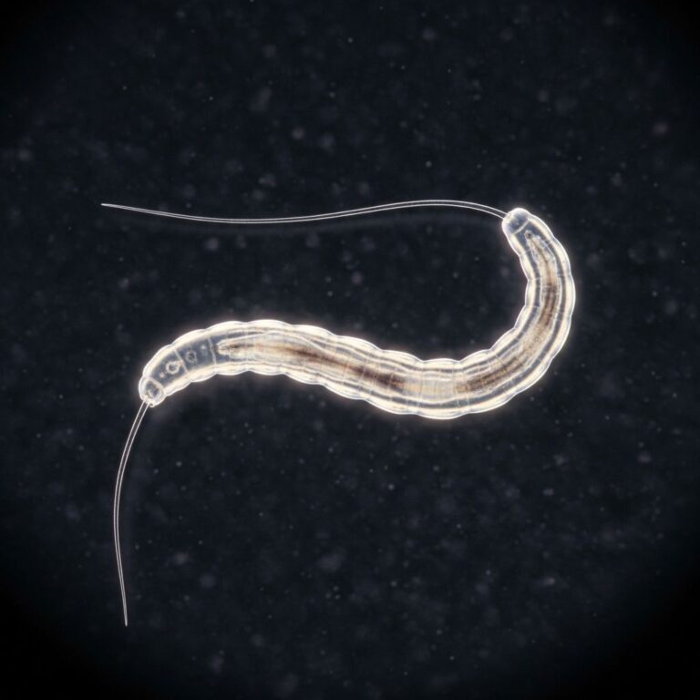
Approximately 50% of keratitis cases are infectious in nature, with bacterial infections accounting for roughly 80% of infectious cases. Bacterial keratitis typically develops rapidly and can cause significant corneal scarring. Viral keratitis, often caused by herpes simplex virus, tends to be more chronic and recurrent. Fungal keratitis is less common but often more difficult to treat. Parasitic keratitis, such as Acanthamoeba keratitis, is rare but particularly aggressive and challenging to manage.
Non-Infectious Keratitis
Non-infectious keratitis includes conditions such as neurotrophic keratopathy, which occurs due to impaired corneal nerve function, and inflammatory keratitis associated with autoimmune conditions. These types often require different treatment approaches than infectious keratitis.
Risk Factors and Causes
Several factors increase your risk of developing keratitis. The most common risk factors include:
Corneal Trauma
Any injury to the cornea, including scratches from foreign objects, chemical burns, or abrasions from contact lenses, can lead to keratitis. Even minor scratches can become infected if not properly treated, making injury prevention crucial for contact lens wearers.
Contact Lens Wear
Contact lens use is a significant risk factor for keratitis, particularly when lenses are not properly cleaned and maintained. Poor hygiene practices, sleeping in contact lenses not designed for overnight wear, and extended wear without proper care dramatically increase infection risk. Users who expose their lenses to tap water or non-sterile solutions face elevated danger of developing keratitis, including serious infections like Acanthamoeba keratitis.
Breakdown of Corneal Epithelium
Conditions that compromise the protective outer layer of the cornea, including dry eye syndrome, reduced tear production, and eyelid problems, significantly increase keratitis susceptibility. The corneal epithelium serves as a barrier against infection, and any disruption to this barrier increases vulnerability.
Other Contributing Factors
Additional risk factors include immunosuppression, diabetes, ocular herpes simplex infection, chemical or thermal burns, and certain medications that affect tear production or corneal sensation. Individuals with compromised immune systems face higher risk of severe infections.
Symptoms of Keratitis
Keratitis typically presents with recognizable symptoms that develop suddenly or gradually depending on the cause. Common symptoms include:
- Eye pain or discomfort that may worsen with light exposure
- Redness of the white part of the eye
- Excessive tearing or discharge
- Blurred or decreased vision
- Foreign body sensation, as if something is in the eye
- Light sensitivity (photophobia)
- Swelling of the eyelid
- Corneal cloudiness or opacity visible to the naked eye in severe cases
The severity and combination of symptoms can vary depending on whether the infection is bacterial, viral, fungal, or parasitic. Acanthamoeba keratitis, for example, is characterized by progressive corneal inflammation, epithelial defects, and ulceration that can lead to significant visual impairment if not promptly treated.
Diagnosis of Keratitis
Accurate diagnosis depends on a careful medical history, comprehensive eye examination, and appropriate diagnostic testing. Eye care professionals use several methods to identify and classify keratitis.
Initial Evaluation
Your eye doctor will review your medical history, including contact lens use, recent eye injuries, and any systemic conditions affecting ocular health. They will ask about symptom onset, progression, and any treatments already attempted.
Slit-Lamp Examination
The slit-lamp microscope allows detailed visualization of the cornea, revealing corneal opacities, infiltrates, and ulceration patterns. This examination helps determine the location, depth, and extent of corneal involvement and is essential for guiding treatment decisions. For Acanthamoeba keratitis specifically, the slit-lamp may reveal a characteristic ring infiltrate.
Corneal Scraping and Cultures
Corneal scraping involves gently collecting a sample from the corneal surface for laboratory analysis. These samples are cultured to identify the causative organism and determine its sensitivity to specific antimicrobial agents. Culture results guide antibiotic selection and improve treatment effectiveness. For Acanthamoeba keratitis, cultures on non-nutrient agar with an Escherichia coli overlay can promote organism growth, though results may require several days.
Additional Diagnostic Tests
Depending on clinical findings, your doctor may recommend:
- Polymerase Chain Reaction (PCR) testing: Provides rapid identification of viral or parasitic organisms
- In Vivo Confocal Microscopy (IVCM): Offers detailed visualization of corneal layers and is particularly valuable for early diagnosis and monitoring treatment response in cases like Acanthamoeba keratitis
- Complete Blood Count (CBC): Evaluates overall infection status and immune response
- HIV testing: Identifies immunosuppression that may affect treatment outcomes
- Autoimmune testing: Determines if non-infectious inflammatory conditions contribute to keratitis
- Anterior Segment Optical Coherence Tomography (AS-OCT): Provides cross-sectional imaging of corneal layers
- Impression cytology: Collects surface cells for microscopic examination
Treatment Options
Treatment approaches vary based on keratitis type, severity, and causative organism. The primary goal is to eliminate infection, reduce inflammation, and preserve vision.
Treatment for Infectious Keratitis
Infections are treated with prescription eye drops containing antimicrobial agents, which may be supplemented by systemic antibiotics, antivirals, or antifungals as needed. For bacterial keratitis, topical fluoroquinolone antibiotics are typically prescribed and often applied frequently throughout the day. For viral keratitis caused by herpes simplex, antiviral eye drops and oral antiviral medications may be used.
Acanthamoeba Keratitis Treatment
Acanthamoeba keratitis demands comprehensive treatment integrating medical management and surgical interventions. Biguanides, such as polyhexamethylene biguanide (PHMB) and chlorhexidine, are typically first-line treatments due to their broad-spectrum antimicrobial properties and effectiveness against Acanthamoeba. Aromatic diamidines serve as alternative or adjunctive agents. Early diagnosis within 14 days of symptom onset generally results in better visual outcomes, while delayed diagnosis is associated with decreased final vision and increased need for surgical intervention.
Supportive Treatments
Beyond antimicrobial agents, treatment may include:
- Pupil-dilating agents: Reduce pain and prevent iris adhesions
- Topical analgesics: Provide pain relief and increase patient comfort
- Corticosteroids: Reduce inflammation, though must be used cautiously in infectious keratitis as premature use before definitive diagnosis can worsen outcomes
- Lubricating eye drops: Maintain corneal moisture and comfort
Surgical Interventions
In advanced or refractory cases, surgical options may be necessary. These include corneal crosslinking to stabilize the cornea and therapeutic keratoplasty (corneal transplantation) for severe scarring or perforation. Emerging therapies such as photodynamic therapy and antimicrobial peptides show promise in enhancing treatment outcomes.
Treatment for Traumatic Keratitis
When keratitis is caused by injury, treatment focuses on supporting natural healing. Antibiotic ointment may be applied to prevent secondary infection while the cornea heals. Most traumatic keratitis cases resolve with appropriate supportive care.
Complications of Untreated Keratitis
Untreated or inadequately managed keratitis can lead to serious complications affecting long-term vision:
- Corneal scarring: Permanent opacification that reduces visual clarity and may require corneal transplantation
- Corneal perforation: Full-thickness corneal rupture leading to corneal collapse and severe vision loss
- Endophthalmitis: Spread of infection to the interior of the eye, a sight-threatening emergency
- Blindness: Permanent vision loss if deep infection causes extensive corneal damage
These serious outcomes emphasize the critical importance of prompt diagnosis and treatment. Even surface keratitis can leave scars that damage vision or cause blindness if infection penetrates deeper than the corneal surface.
Prevention Strategies
Preventing keratitis involves protecting your eyes from injury and maintaining proper lens hygiene if you wear contacts:
- Contact lens care: Clean and disinfect lenses daily using only approved solutions, never tap water
- Proper lens wear: Follow prescribed wear schedules and replace lenses as recommended
- Eye protection: Wear protective eyewear during activities with injury risk
- Maintain eye health: Treat dry eye syndrome and manage underlying conditions
- Hygiene practices: Wash hands before handling contact lenses
- Avoid chemical exposure: Protect eyes from harsh chemicals and irritants
Prognosis and Recovery
Keratitis is usually easy to treat and clears up quickly when managed appropriately. However, recovery depends on several factors including infection severity, organism type, timing of diagnosis, and treatment adherence. Patients receiving treatment within 14 days of symptom onset generally experience better visual outcomes, particularly in Acanthamoeba keratitis cases. With early intervention and proper management, most people recover their vision completely without lasting damage.
When to Seek Medical Attention
Contact an eye care professional immediately if you experience:
- Sudden eye pain or significant discomfort
- Redness that develops rapidly
- Vision changes or blurriness
- Excessive tearing or discharge
- Light sensitivity
- Foreign body sensation that doesn’t resolve
- Eye symptoms following trauma or contact lens problems
Keratitis is an ocular emergency requiring prompt professional evaluation to prevent permanent vision loss.
Frequently Asked Questions
Q: Can keratitis cause permanent blindness?
A: Yes, untreated keratitis or delayed treatment can lead to corneal scarring and permanent vision loss or blindness, especially when infection penetrates deep into the cornea. Early diagnosis and treatment are essential to prevent these complications.
Q: Is keratitis contagious?
A: Infectious keratitis caused by bacteria, viruses, or parasites can potentially spread through contact, particularly viral and some bacterial forms. However, proper hygiene and contact lens care significantly reduce transmission risk.
Q: How long does keratitis take to heal?
A: Recovery time varies depending on keratitis type and severity. Mild surface keratitis may resolve within days to weeks with appropriate treatment, while severe infections or those affecting deeper corneal layers may require weeks to months for complete healing.
Q: Can I wear contact lenses if I have keratitis?
A: No, contact lens wear should be discontinued immediately if keratitis is suspected or confirmed. Lenses can trap infection against the cornea and delay healing. Always consult your eye doctor before resuming contact lens use after keratitis.
Q: What is Acanthamoeba keratitis?
A: Acanthamoeba keratitis is a rare but severe corneal infection caused by a free-living amoeba commonly found in water. It is most prevalent among contact lens wearers with poor hygiene practices and can lead to significant vision loss if not promptly diagnosed and treated with specialized antimicrobial therapy.
Q: Are there effective treatments for keratitis?
A: Yes, keratitis is usually easy to treat with appropriate medications including topical antimicrobial eye drops, systemic antibiotics or antivirals when needed, and supportive care. Treatment success depends on early diagnosis, proper organism identification, and adherence to prescribed therapy.
References
- Keratitis – Symptoms, diagnosis and treatment — BMJ Best Practice US. 2025. https://bestpractice.bmj.com/topics/en-us/561
- A Comprehensive Review on Acanthamoeba Keratitis: An Overview — National Center for Biotechnology Information, U.S. National Library of Medicine. 2024. https://pmc.ncbi.nlm.nih.gov/articles/PMC11424229/
- Keratitis: What to Do to Save Your Sight — WebMD Eye Health. 2025. https://www.webmd.com/eye-health/keratitis-facts
- Diagnosis and Treatment of Neurotrophic Keratopathy — Johns Hopkins School of Medicine CME. 2025. https://hopkinscme.cloud-cme.com/assets/hopkinscme/Presentations/28879/28879.pdf
- Ocular Keratitis — Johns Hopkins ABX Guide. 2025. https://www.hopkinsguides.com/hopkins/view/Johns_Hopkins_ABX_Guide/540395/all/Ocular_Keratitis
- Acanthamoeba — Johns Hopkins ABX Guide. 2025. https://www.hopkinsguides.com/hopkins/view/Johns_Hopkins_ABX_Guide/540002/all/Acanthamoeba
Similar Articles


Read full bio of medha deb