Keratosis Pilaris Images: Comprehensive Clinical Photo Atlas
Explore detailed images and clinical insights into keratosis pilaris, a common skin condition causing rough, bumpy patches.

Keratosis pilaris (KP), often called “chicken skin,” is a prevalent dermatological condition characterized by small, rough bumps due to keratin buildup blocking hair follicles. This image gallery provides a visual atlas of KP manifestations across different body sites, skin types, and clinical variants, aiding in diagnosis and patient education.
What is keratosis pilaris?
Keratosis pilaris develops when excess keratin—a protective skin protein—forms scaly plugs that obstruct hair follicle openings, resulting in patches of rough, bumpy skin. This harmless condition affects up to 40-50% of adults and 50-80% of adolescents, often running in families due to genetic factors. It is more noticeable in winter or dry climates when skin hydration decreases, exacerbating the follicular hyperkeratosis.
KP cannot be cured but improves with age, frequently resolving by the third decade of life. Management focuses on moisturization and gentle exfoliation to smooth the skin texture.
Classic keratosis pilaris
The hallmark of classic KP includes numerous follicular keratotic plugs presenting as tiny, spiny red-based papules, predominantly on the upper outer arms and thighs. These bumps resemble sandpaper and may have a background erythema.
- Arm presentation: Close-up views reveal 1-2 mm follicular papules with central keratin plugs on erythematous bases, coalescing into rough plaques.
- Thigh involvement: Similar bumps scatter across anterolateral thighs, often symmetric and more pronounced post-bath due to trapped moisture.
Image captions typically highlight: ‘Keratosis pilaris on upper arms showing classic follicular hyperkeratosis with perifollicular erythema.’
Keratosis pilaris rubra
This variant features more intense background redness surrounding the keratotic follicles, creating a vivid “goosebump” appearance. It is particularly common in lighter skin types and may itch mildly.
- Images depict diffuse erythema on cheeks or arms dotted with white-topped spines.
- Patches on buttocks show mottled red discoloration with pinpoint keratoses.
Visuals emphasize the rubra subtype’s florid inflammation: ‘KP rubra on the cheek with prominent perifollicular redness and subtle scaling.’
Keratosis pilaris atrophicans
A rarer, progressive form where chronic inflammation leads to perifollicular atrophy, resulting in pale, depressed macules amid keratotic plugs. This can mimic scarring and is associated with atrophicans faciei variant on cheeks.
- Facial images show ivory-colored atrophic patches with follicular spines on cheeks.
- Arm views reveal hypopigmented macules surrounding resolved follicles.
Captions note: ‘Keratosis pilaris atrophicans faciei demonstrating follicular atrophy and background pallor.’
KP of the cheeks
Infants and young children often present with cheek-predominant KP, featuring sandpaper-like roughness without significant redness. This resolves spontaneously but can persist.
- Close-ups illustrate monomorphic 1 mm papules blending seamlessly across malar cheeks.
- Comparison images contrast normal vs. KP cheeks under raking light.
KP on buttocks
Buttock involvement yields clustered bumps in a “popcorn” texture, worsened by friction from clothing. Images capture posterior thigh-buttock extension.
- Patches show hyperpigmented plaques in darker skin types.
- Lateral views highlight extension to flanks.
Unusual sites
While arms and thighs dominate, KP can appear on the forearms, calves, flanks, and rarely palms or soles. Images document these ectopic presentations.
- Forearm: Linear arrangement along extensors.
- Deltoids and sacral area: Scattered discrete papules.
KP in darker skin types
In skin of color, KP manifests with hyperpigmented follicular papules rather than red bases, leading to post-inflammatory pigment changes. Images showcase brown-to-black spiny lesions on thighs.
- Thighs: Confluent hyperkeratotic plaques with pigment tropism.
- Arms: Ashy, scaly follicular accentuation.
This variant risks keloidal scarring if aggressively treated.
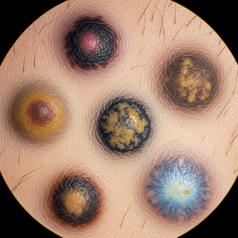
Close-up of individual lesions
Magnified views reveal the pathognomonic triad: central keratin horn atop a hair stub, surrounded by erythema, within a dilated follicle. These 1-2 mm lesions are firmly attached and non-tender.
- Stereomicroscopy-like images detail plug extrusion post-exfoliation.
- Histology correlates: orthokeratotic plugging of follicles.
Dermoscopy findings
Dermoscopic evaluation aids differentiation from acne or folliculitis, showing white annular structures (hair casts) with peripheral red loops.
- Patterns: targetoid white dots amid structureless red.
- Vascular: comma-shaped vessels in rubra subtype.
Histopathology
Microscopic sections confirm absent inflammation but marked hyperkeratosis plugging the infundibulum. Follicular ostia appear widened with retained corneocytes.
- H&E stain: orthokeratosis, absent granular layer disruption.
- Images compare normal vs. KP follicles.
Treatment effects – before and after
Sequential images demonstrate improvement with topical therapies: urea/lactic acid creams reduce plug size within 4-6 weeks, while lasers smooth texture.
- Pre-treatment: Dense sandpaper texture on arms.
- Post 12 weeks urea 20%: 70% clearance, residual faint erythema.
- Laser post-PDT: Reduced discoloration in skin of color.
Clinical variants table
| Variant | Key Features | Common Sites | Image Characteristics |
|---|---|---|---|
| Classic KP | Follicular papules, mild erythema | Arms, thighs | Sandpaper roughness, red-based spines |
| KP Rubra | Intense redness | Cheeks, arms | Goosebump-like with background flush |
| KP Atrophicans | Atrophic macules | Face, arms | Pale depressions amid keratoses |
| Cheek KP (infantile) | Smooth roughness | Cheeks | Monomorphic papules, no scaling |
| Dark skin KP | Hyperpigmented plugs | Thighs, buttocks | Brown follicular accentuation |
Frequently Asked Questions
Is keratosis pilaris permanent?
No, KP often improves or resolves by age 30, though it can persist lifelong in milder forms. Consistent moisturizing prevents flare-ups.
Does KP itch or hurt?
Rarely painful, but dryness can cause itchiness, especially in rubra variant. Avoid scratching to prevent pigmentary changes.
Can I exfoliate KP skin?
Gentle chemical exfoliants (lactic acid, urea) yes; harsh scrubs no, as they worsen inflammation.
Is KP contagious?
No, it’s a genetic keratinization disorder, not infectious.
When to see a dermatologist?
If OTC treatments fail, significant redness/itch occurs, or for laser options in cosmetic concerns.
Management Summary
Daily fragrance-free moisturizers with exfoliants (urea 10-20%, lactic acid 12%, salicylic acid 2%) form the cornerstone. Topical retinoids and brief steroids address inflammation. Procedures like microdermabrasion or pulsed dye laser offer advanced smoothing.
References
- Keratosis Pilaris — Loma Linda University Health. 2023. https://lluh.org/conditions/keratosis-pilaris
- Keratosis pilaris – treatment, causes and symptoms — Healthdirect (Australian Government). 2024-01-15. https://www.healthdirect.gov.au/keratosis-pilaris
- Keratosis Pilaris Treatment NYC — Manhattan Dermatology Specialists. 2023. https://www.manhattandermatologistsnyc.com/procedures/keratosis-pilaris-treatment/
- Understanding Keratosis Pilaris: Causes, Symptoms, and Treatments — Dr. Snyder Dermatology. 2024. https://drsnyder.com/understanding-keratosis-pilaris-causes-symptoms-and-treatments/
- Keratosis pilaris – Symptoms and causes — Mayo Clinic. 2024-06-12. https://www.mayoclinic.org/diseases-conditions/keratosis-pilaris/symptoms-causes/syc-20351149
- Keratosis pilaris: Diagnosis and treatment — American Academy of Dermatology (AAD). 2024. https://www.aad.org/public/diseases/a-z/keratosis-pilaris-treatment
Similar Articles


Read full bio of Sneha Tete