Laboratory Tests For Fungal Infection: A Practical Guide
Comprehensive guide to essential lab tests for diagnosing fungal infections of skin, hair, nails, and beyond.

Skin, hair, and nail tissue are collected for microscopy and culture (mycology) to establish or confirm the diagnosis of a fungal infection. These tests are crucial for identifying dermatophytes, yeasts, and other fungi causing superficial, subcutaneous, or systemic infections.
Introduction
Fungal infections affect the skin, hair, nails, and sometimes deeper tissues or systemic organs. While clinical appearance often suggests a fungal cause, laboratory confirmation is essential, especially for chronic, severe, or atypical cases considering systemic therapy. Common pathogens include dermatophytes (e.g., Trichophyton, Microsporum, Epidermophyton), yeasts like Candida and Malassezia, and moulds. Accurate diagnosis guides targeted antifungal therapy, preventing unnecessary treatments and resistance.
Diagnosis relies on a combination of direct microscopy for rapid detection, culture for species identification, and increasingly, molecular methods for speed and sensitivity. Negative results do not always exclude infection due to sampling issues or low fungal load.
Specimen Collection
Proper specimen collection is critical for reliable results. Specimens should be gathered from the most active lesion sites, preferably before starting antifungal treatment, to maximize yield. Quantity matters—collect ample material for optimal testing.
Transport specimens in sterile containers or black paper envelopes to the mycology lab promptly. For pustules, include a swab to rule out secondary bacterial infection.
- Skin scrapings: Use a sterile blade or curette to gently scrape scales from the active border of lesions (e.g., ring edge in tinea). Clean the site with alcohol first. Avoid recent creams or scrubbing, which reduce yield.
- Hair: Pluck hairs with forceps, including roots and sheaths, from infected areas. For tinea capitis, select fluorescing hairs under Wood’s lamp if applicable.
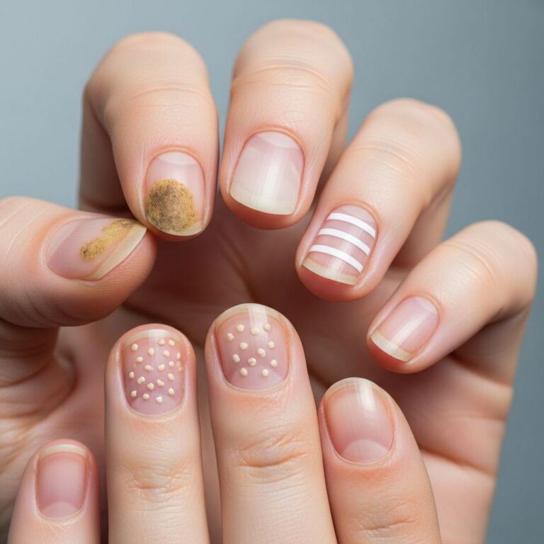
- Nails: Clip proximal diseased nail, scrape subungual debris. Target the junction of normal and abnormal nail for best results (see Figure 1 description below).
- Other: Biopsies for subcutaneous/deep infections; pus, sputum, or tissue for systemic cases.
Wood’s Lamp Examination: Expose to long-wavelength UV light; some tinea capitis hairs fluoresce green-yellow due to pteridine production by Microsporum species. Not diagnostic alone but aids sampling.
Figure 1 (Adapted): Nail specimens from proximal undersurface, clipping abnormal areas and scraping debris.
Collection is generally painless, though subungual scraping may cause mild discomfort. Repeat if initial tests are negative but suspicion persists.
Direct Microscopy
Microscopy provides rapid results (minutes) by visualizing fungal elements in specimens treated to dissolve keratin.
Process: Mount specimen in 10-20% potassium hydroxide (KOH) to clear keratinocytes, sometimes with blue/black ink or calcofluor white for contrast under fluorescence microscopy. Examine under light or fluorescence scope.
- Dermatophytes: Septate branching hyphae (2-10 µm wide), arthroconidia (spores), mosaic patterns in nails.
- Yeasts: Blastospores, pseudohyphae (e.g., Candida); ‘spaghetti and meatballs’ appearance; Malassezia as short hyphae with round yeasts (‘perifolliculitis’).
- Moulds/Dimorphics: Varied hyphae, spores; special stains like PAS or GMS for biopsies.
Fungal elements may be scarce in inflamed tissue, yielding false negatives (sensitivity ~60-80%). Does not identify species.
In histopathology (for biopsies), PAS, GMS, or Gram stains highlight fungi in tissue context, assessing invasion/inflammation.
Culture
Culture confirms infection and identifies species/antifungal susceptibility, but takes 1-4 weeks at 25-30°C.
Inoculate on Sabouraud dextrose agar (SDA) with cycloheximide (inhibits contaminants) and chloramphenicol (antibacterial). Omit cycloheximide for moulds. Observe colony morphology, microscopy (lactophenol cotton blue mount) for macro/microconidia, spirals, etc.
False negatives from poor sampling, prior antifungals, or non-viable fungi. Positive cultures may represent colonization, not infection (e.g., in psoriasis).
| Fungus Type | Key Culture Features | Common Sites |
|---|---|---|
| Dermatophytes | Velvety/powdery colonies; septate hyphae, conidia | Skin, hair, nails |
| Candida spp. | Creamy, pasty; pseudohyphae, buds | Mucosa, nails |
| Malassezia | Lipophilic; requires oils; bottle-shaped yeasts | Pityriasis versicolor |
Blood Tests
Irrelevant for superficial infections; useful for subcutaneous/systemic mycoses (e.g., sporotrichosis, histoplasmosis).
- Antigen Detection: Galactomannan (Aspergillus), beta-D-glucan (multiple fungi); serum/cerebrospinal fluid.
- Antibodies: IgG/IgM for endemic fungi (e.g., coccidioidomycosis); less specific.
- PCR: Blood/tissue for disseminated infection.
Combine with imaging/culture for systemic diagnosis.
Molecular Biology Techniques
PCR and sequencing offer high sensitivity/specificity, results in 24 hours, ideal for treatment/epidemiology. Targets ITS/rRNA genes.
- Advantages: Detects non-culturable/viable fungi; species ID; from FFPE tissue (though less reliable).
- IHC/ISH: Tissue stains for antigens/nucleic acids; confirms morphology.
- Emerging: MALDI-TOF MS for rapid ID from cultures.
PCR superior to microscopy/culture; gold standard when histology + PCR. Cost limits routine use, but expanding.
Frequently Asked Questions (FAQs)
Q: When should lab tests be requested for suspected fungal infection?
A: For chronic/severe cases, uncertain diagnosis, systemic therapy consideration, treatment failure, or outbreaks.
Q: Can a negative microscopy or culture rule out fungal infection?
A: No—false negatives common due to low burden, prior treatment, or sampling error. Repeat if suspicion high.
Q: How long do culture results take?
A: 1-4 weeks; most positives in 1-2 weeks. PCR faster (24h).
Q: Are blood tests useful for skin fungal infections?
A: No, for superficial only. Yes for deep/systemic.
Q: What’s the role of biopsies in fungal diagnosis?
A: Essential for subcutaneous/unusual presentations; enables histology + IHC/PCR.
This comprehensive approach ensures precise diagnosis, optimizing patient outcomes in fungal infections.
References
- Laboratory tests for fungal infections — DermNet NZ. 2023-06-15. https://dermnetnz.org/topics/laboratory-tests-for-fungal-infection
- Collecting specimens for the investigation of fungal infections — bpac.org.nz. 2011-03-01. https://bpac.org.nz/BT/2011/March/fungal-infections.aspx
- Dermatopathology and the Diagnosis of Fungal Infections — PMC (NCBI). 2023-06-12. https://pmc.ncbi.nlm.nih.gov/articles/PMC10282148/
- Fungal Nail Infections — DermNet NZ. 2023-08-20. https://dermnetnz.org/topics/fungal-nail-infections
- Mycology – Fungal skin infections — DermNet NZ. 2023-05-10. https://dermnetnz.org/cme/fungal-infections/mycology
Similar Articles


Read full bio of Sneha Tete