Laugier-Hunziker Syndrome: Clinical Features and Management
Understanding benign pigmentary disorder affecting oral mucosa, nails, and skin.

Laugier-Hunziker Syndrome: Overview and Clinical Characteristics
Laugier-Hunziker syndrome is a rare, benign pigmentary disorder characterized by the distinctive expression of pigmentation across mucosal, nail, and acral sites. This condition primarily affects adults and is marked by the appearance of dark spots (hyperpigmented macules) on the oral mucosa and lips. Despite its rarity, understanding this syndrome is essential for healthcare professionals, particularly dentists and dermatologists, as it may be confused with more serious pigmentary conditions.
The condition was first documented by Laugier and Hunziker, who reported a series of five patients with hyperpigmented macules of the mouth and lips, with two of them also presenting with pigment changes of the fingernails. Since that initial description, approximately 100 cases have been reported in medical literature. The syndrome affects individuals across a wide age range, from the second to the ninth decade of life, with both men and women equally susceptible.
Clinical Features and Presentation
The main clinical manifestation of Laugier-Hunziker syndrome is the appearance of pigmentation on specific oral regions. The buccal mucosa (inner lining of the cheeks) and lips, particularly the lower lip, are the most commonly affected areas. However, pigmentation can also extend to other intraoral sites, including:
- Gingiva (gums)
- Tongue
- Roof of the mouth (hard palate)
- Posterior pharyngeal mucosa
The oral lesions appear as light-brown to brown-black macules, typically measuring between 0.1 to 0.5 cm in diameter. These pigmented areas present as well-demarcated spots that may vary in size and shape.
Nail Involvement
Oral and nail involvement occurs concurrently in approximately 60% of cases. Nail pigmentation manifests primarily through longitudinal melanonychia, which presents as linear, pigmented streaks running along the length of the nail. This feature is present in the majority of cases and is permanent in nature. The nail pigmentation can appear in the following patterns:
- Single longitudinal streaks on one or multiple nails
- Multiple pigmented lines on the same nail
- Involvement of both fingernails and toenails
- Hutchinson sign, defined as the extension of pigment onto the proximal nail fold
Fingernail and toenail pigmentation is observed in half to two-thirds of cases and represents a distinctive feature that aids in diagnosis.
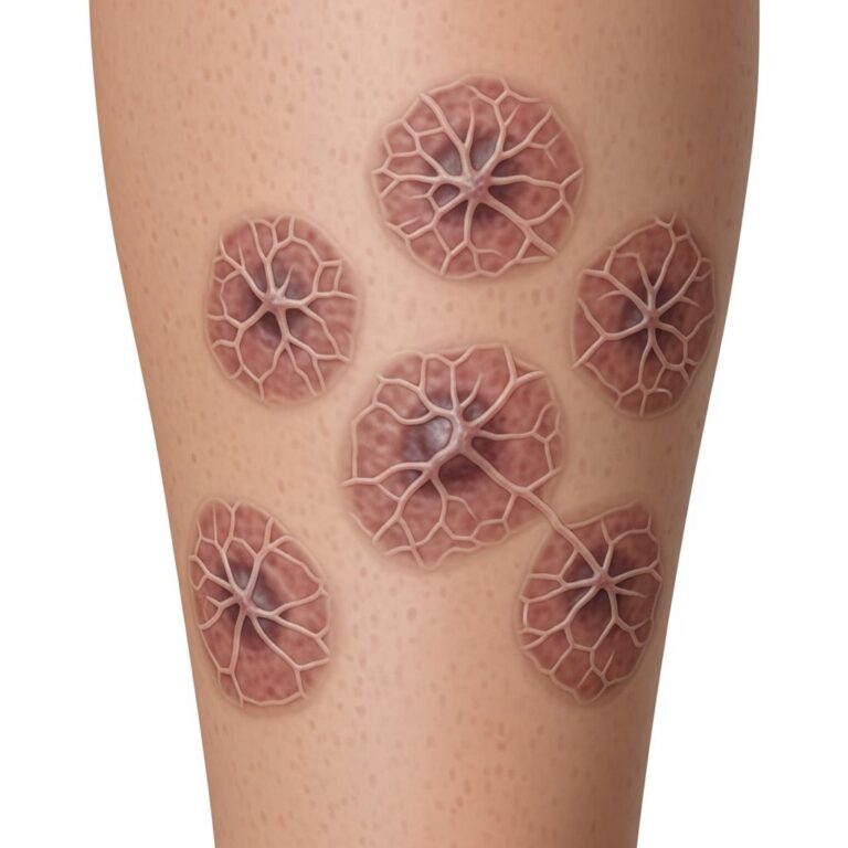
Skin Manifestations
Hyperpigmented macules on the skin sometimes appear on various body sites, including:
- Neck and abdomen
- Fingers and palms
- Soles of the feet
- Genital region (vulva, penis)
- Anal mucosa
- Interdigital areas (spaces between fingers and toes)
- Pretibial area (front of the lower leg)
- Conjunctiva (covering of the eye)
The distal aspects of the fingers and toes frequently display hyperpigmented macules similar to those found on the oral mucosa.
Age of Onset and Inheritance Pattern
Laugier-Hunziker syndrome typically manifests during early or mid-adult life. Patients often report that lesions appeared several years prior to seeking medical consultation, as the primary concern is usually cosmetic rather than health-related.
The inheritance pattern of this syndrome remains variable and somewhat controversial. The condition has been reported as sporadic in most cases, but case reports have included related family members, suggesting possible autosomal dominant inheritance in some families. However, the precise genetic basis and inheritance mechanism remain unclear, with some cases showing associations with potential genetic disorders.
Histopathological Features
Microscopic examination of mucosal macules reveals important diagnostic characteristics. The histopathology typically shows:
- Normal numbers and appearance of melanocytes, although some cases describe increased melanocyte numbers
- Accumulation of pigment in the basal epidermal layer
- Presence of melanophages in the reticular dermis
- Large numbers of mature melanosomes in the stratum basale on electron-microscopy examination
- Absence of nevus cells
The current presumed etiology is that elevated activity of melanocytes leads to increased melanosome formation, resulting in melanin accumulation in the basal mucosal layer, while maintaining a normal number and morphologic appearance of melanocytes.
Associated Conditions and Complications
While Laugier-Hunziker syndrome is fundamentally a benign condition with no association with systemic diseases, rare case reports have described concurrent presentations with other conditions, including:
- Esophageal melanocytosis
- Actinic lichen planus
- Hypocellular marrow and thrombocytopenia
- Invasive melanoma
- Lupus erythematosus
However, the relationships between Laugier-Hunziker syndrome and these conditions have not been well established, and they may represent coincidental findings rather than true associations.
Differential Diagnosis
Accurate diagnosis is crucial to differentiate Laugier-Hunziker syndrome from other conditions that present with oral pigmentation. The primary differential diagnoses include:
| Condition | Distinguishing Features |
|---|---|
| Drug-Induced Pigmentation | History of medication use (phenytion, antimalarials, clofazimine, zidovudine, phenothiazine); resolves upon drug discontinuation |
| Addison Disease | Associated systemic symptoms; abnormal adrenal function tests; generalized hyperpigmentation |
| Albright Syndrome | Café-au-lait spots; endocrine abnormalities; developmental bone disease |
| Peutz-Jeghers Syndrome (PJS) | Congenital or early childhood onset; associated gastrointestinal polyps; family history of GI complications |
| Physiological Pigmentation | Solitary lesions; increases with age; absence of nail and systemic involvement |
| Melanotic Macules | Usually present as solitary areas of pigmentation; limited to oral mucosa |
| Oral Mucosal Melanoma | New lesion from apparently normal mucosa; rapid progression; may have 30-50% persistent pigmentation |
Key diagnostic features that support Laugier-Hunziker syndrome include the presence of multiple areas of oral pigmentation accompanied by nail and skin involvement, absence of systemic symptoms, unremarkable medical and medication history, and benign clinical course.
Diagnosis and Clinical Evaluation
The diagnosis of Laugier-Hunziker syndrome is primarily clinical, based on characteristic presentation of pigmentation pattern across multiple sites. A thorough medical history should establish:
- Age of onset and duration of lesions
- Progression pattern (stable versus progressive darkening)
- Absence of systemic symptoms
- Current and past medication use
- Family history of similar conditions
Physical examination should systematically assess the oral cavity, nails, and skin for characteristic pigmentation patterns. Dermoscopy or dermoscopic evaluation may help confirm the benign nature of lesions. Laboratory abnormalities are typically absent or minimal, and routine laboratory workup is generally unnecessary unless systemic symptoms are present, warranting investigation for alternative diagnoses.
Treatment and Management
No active treatment is required for Laugier-Hunziker syndrome. This is a benign, non-progressive condition that does not pose health risks and has no association with systemic diseases. Management focuses on appropriate diagnosis and patient reassurance.
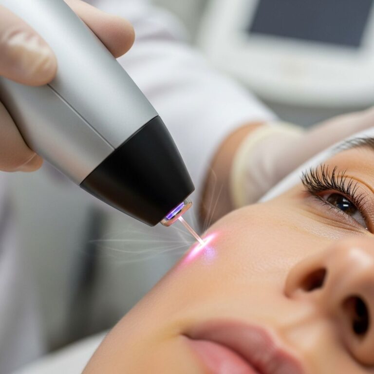
However, treatment options are available for patients who experience cosmetic distress. Cosmetically disfiguring or bothersome lesions may be treated with laser therapy, which can help reduce the visibility of pigmented areas. The selection of specific laser wavelengths depends on the depth and type of pigmentation, with consultation from a dermatologist recommended for optimal outcomes.
Regular follow-up and monitoring are recommended to ensure the stability of lesions and to promptly identify any unusual changes that might warrant further investigation.
Patient Counseling and Prognosis
Patients diagnosed with Laugier-Hunziker syndrome should receive clear explanation regarding the benign nature of their condition. Key points in patient education include:
- The condition is benign and non-malignant
- Pigmentation typically remains stable or progresses very slowly
- There is no association with systemic disease
- The primary concern is cosmetic appearance rather than health risk
- Treatment is optional and purely for cosmetic purposes
- Long-term prognosis is excellent with no serious complications
The prognosis for Laugier-Hunziker syndrome is excellent. Patients experience minimal complications beyond the cosmetic aspect of their condition. Most individuals seek consultation after several years of pigmentation, often due to cosmetic concerns rather than health-related issues.
Frequently Asked Questions
Q: Is Laugier-Hunziker syndrome hereditary?
A: The inheritance pattern is variable. While most cases appear sporadic, some case reports have documented familial involvement suggesting possible autosomal dominant inheritance in certain families. However, the genetic basis remains unclear.
Q: Can Laugier-Hunziker syndrome turn into melanoma?
A: Laugier-Hunziker syndrome is a benign condition with no malignant potential. While rare cases of invasive melanoma have been reported concurrently with the syndrome, this represents coincidental occurrence rather than malignant transformation.
Q: Is treatment necessary for Laugier-Hunziker syndrome?
A: No active treatment is required as the condition is benign. Treatment with laser therapy is optional and recommended only for patients who find the pigmentation cosmetically bothersome.
Q: How is Laugier-Hunziker syndrome diagnosed?
A: Diagnosis is primarily clinical, based on characteristic presentation of multiple hyperpigmented macules on oral mucosa, nails, and skin, combined with unremarkable medical history and absence of systemic symptoms. Histopathology can confirm the diagnosis if needed.
Q: What is the difference between Laugier-Hunziker syndrome and Peutz-Jeghers syndrome?
A: While both conditions present with oral pigmentation, Peutz-Jeghers syndrome presents with congenital or early childhood onset pigmentation and is associated with gastrointestinal polyps and complications, whereas Laugier-Hunziker syndrome has adult onset and no systemic associations.
Q: Will the pigmentation spread or worsen over time?
A: Pigmentation in Laugier-Hunziker syndrome typically remains stable or progresses very slowly. Most patients report their lesions have been present for several years with minimal change, though some cases show gradual darkening.
References
- Laugier Hunziker Syndrome — eScholarship.org. 2024. https://escholarship.org/uc/item/59q2q20w
- Laugier-hunziker Syndrome — DermNet. 2024. https://dermnetnz.org/topics/laugier-hunziker-syndrome
- Laugier-Hunziker Syndrome: A Rare Cause of Oral Mucosa Pigmentation — National Institutes of Health, PubMed Central. 2023. https://pmc.ncbi.nlm.nih.gov/articles/PMC10411268/
- Laugier-Hunziker Syndrome — StatPearls, NCBI Bookshelf. 2024. https://www.ncbi.nlm.nih.gov/books/NBK534300/
- Laugier-Hunziker Syndrome: A Clinical Review of Six Cases — Clinical and Experimental Dermatology, Oxford Academic. 2024. https://academic.oup.com/ced/article/15/2/111/6629047
Similar Articles


Read full bio of medha deb