Leg Ulcers: Expert Guide To Diagnosis, Treatment & Prevention
Comprehensive guide to causes, diagnosis, and evidence-based management of chronic leg ulcers for optimal healing.

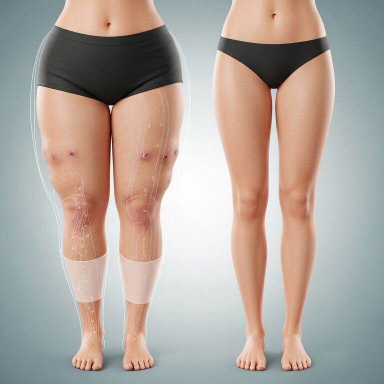
Leg ulcers are defined as full-thickness skin loss on the lower leg persisting for more than 3 months, often resulting from venous insufficiency, arterial disease, or other underlying conditions. Effective management requires accurate diagnosis, addressing the underlying cause, and comprehensive wound care to promote healing and prevent recurrence.
What is a leg ulcer?
A
leg ulcer
represents a chronic breakdown of the skin on the lower leg, typically below the knee, characterised by full-thickness loss of the epidermis and dermis. These ulcers fail to heal within 6 weeks and are considered chronic after 3 months. They affect approximately 1% of the adult population, with prevalence increasing to 3-4% in those over 65 years, posing significant morbidity and healthcare burden.Chronic leg ulcers arise from impaired wound healing due to underlying vascular, neuropathic, or inflammatory pathologies. The most common type is the venous ulcer, accounting for 70-90% of cases, followed by arterial (10-20%) and mixed aetiology. Healing requires a moist environment, debridement of necrotic tissue, infection control, and correction of contributing factors like oedema and malnutrition.
Who gets leg ulcers?
Leg ulcers predominantly affect older adults, with risk factors including:
- Age over 60 years
- Obesity and sedentary lifestyle
- Previous deep vein thrombosis or leg injury
- Family history of venous disease
- Peripheral vascular disease or diabetes
- Immobility or chronic oedema
Venous ulcers are more common in women, while arterial ulcers affect men equally or more. Incidence rises sharply after age 75, impacting quality of life through pain, immobility, and social isolation.
What causes leg ulcers?
The primary causes are vascular insufficiencies disrupting normal wound repair. Key aetiologies include:
Venous leg ulcers
Result from chronic venous hypertension due to valvular incompetence in superficial, deep, or perforator veins, or venous obstruction. This leads to ambulatory venous hypertension, capillary damage, fibrin deposition, and lipodermatosclerosis. Ambulatory venous pressure exceeds 30 mmHg (normal <20 mmHg), causing tissue hypoxia and ulceration, typically at the gaiter area (medial malleolus).
Arterial leg ulcers
Caused by peripheral arterial occlusive disease reducing blood supply (ankle-brachial pressure index, ABPI <0.9). Atherosclerosis leads to ischaemic necrosis, often on toes, feet, or lateral malleoli. Night pain with leg elevation is characteristic due to dependent perfusion.
Diabetic/neuropathic ulcers
Occur in diabetic patients with peripheral neuropathy and microvascular disease, typically over pressure points like the heel or metatarsal heads. Loss of protective sensation leads to repetitive trauma and infection.
Other causes
- Pressure ulcers: From prolonged immobility over bony prominences.
- Pyoderma gangrenosum: Inflammatory, painful ulcers with undermined edges.
- Malignancy: Basal or squamous cell carcinomas arising in chronic wounds (Marjolin ulcer).
- Drugs: Hydroxyurea-induced ulcers in myeloproliferative disorders.
- Infections: Rarely, bacterial, fungal, or mycobacterial.
Mixed arterial-venous disease complicates 20% of cases, requiring vascular assessment before compression therapy.
Clinical features of leg ulcers
Diagnosis relies on history, examination, and investigations. Venous ulcers present as shallow, exudative lesions with irregular borders, granulating base, and surrounding haemosiderin pigmentation, varicose eczema, and lipodermatosclerosis. Pain is mild, relieved by elevation.
Arterial ulcers are deep, ‘punched-out’, with pale/necrotic bases, minimal exudate, and severe ischaemic pain worsened by elevation. Pulses are absent, skin atrophic and hairless.
| Feature | Venous Ulcer | Arterial Ulcer | Neuropathic Ulcer |
|---|---|---|---|
| Location | Medial malleolus | Toes, lateral malleolus | Pressure points (heel) |
| Appearance | Shallow, exudative | Deep, necrotic | Callused edges |
| Pain | Mild, < elevation | Severe, > elevation | Painless |
| ABPI | >0.8 | <0.8 | Variable |
Diagnosis of leg ulcers
History: Onset, trauma, comorbidities, medications.
Examination: Ulcer characteristics, limb volume, oedema, pulses, skin changes, ABPI using Doppler ultrasound (>0.9 normal, 0.6-0.9 mixed, <0.5 severe ischaemia). Compression contraindicated if ABPI <0.8.
Investigations:
- Duplex ultrasound for venous reflux/obstruction.
- Full blood count, glucose, inflammatory markers.
- Biopsy if atypical, non-healing, or suspicious for malignancy.
- Microbiology swabs only if clinical infection (not colonisation).
Biofilm presence in chronic ulcers delays healing; diagnosed clinically by persistent slough despite debridement.
Complications of leg ulcers
- Infection: Cellulitis, osteomyelitis; biofilm protects organisms.
- Malignancy: 2-10% risk in chronic ulcers.
- Recurrence: 60-70% within 1 year without compression hosiery.
- Pain and immobility: Leading to depression, DVT risk.
- Amputation: In severe arterial/diabetic cases.
Management of leg ulcers
Treatment addresses the cause, optimises healing environment, and prevents recurrence. Multidisciplinary input from dermatology, vascular surgery, and wound specialists is ideal.
Treat the cause
- Venous: Compression therapy heals 40-70% within 12 weeks if ABPI >0.8.
- Arterial: Revascularisation (angioplasty, bypass), avoid compression.
- Diabetic: Offloading, glycaemic control.
Compression therapy
The cornerstone for venous ulcers. Options include:
- Multi-layer bandaging: 4-layer systems (e.g., Profore™) providing 40 mmHg at ankle, graduating proximally.
- Hosiery: Class 1-3 (14-40 mmHg); difficult over ulcers.
- Unna boot: Zinc-impregnated gauze hardened with elastic bandage.
- Devices: Intermittent pneumatic compression.
Padding protects bony areas; recheck ABPI weekly initially. Lifelong class 2 hosiery prevents recurrence.
Debridement
Removes necrotic tissue, biofilm, and slough to convert chronic to acute wounds. Methods:
- Autolytic: Occlusive dressings.
- Mechanical: Wet-dry, larvae (maggot) therapy.
- Enzymatic: Ointments.
- Sharp/surgical: Most effective.
Surfactant irrigants disrupt biofilm.
Wound dressings and occlusion
Maintain moist healing environment; change frequency minimised to preserve granulation. Categories:
- Absorbent: Foams, alginates for exudate.
- Non-adherent: Silicones, hydrocolloids.
- Antimicrobial: Silver, honey for infection.
- Occlusive: Promote autolysis.
Hydrocolloids useful for clean ulcers; weekly changes suffice.
Treat infection
Distinguish colonisation (biofilm, slough) from infection (erythema, fever). Systemic antibiotics for cellulitis; topical avoided due to resistance. Swabs unreliable.
Accelerate healing
- Nutrition: Protein, vitamin C, zinc, iron supplements if deficient.
- Leg elevation, exercise to enhance calf pump.
- Skin grafts: Pinch, split-thickness for large clean ulcers post-shave excision of lipodermatosclerosis.
Advanced therapies: Growth factors, skin substitutes (e.g., Apligraf® for venous ulcers), extracellular matrices require further evidence.
Surgery
Endovenous ablation, stripping for superficial reflux; skin grafting for non-healers. Flaps for complex defects.
Prevention of leg ulcers
Target at-risk patients with:
- Weight management, exercise.
- Early treatment of varicose veins.
- Compression hosiery post-healing.
- Emollients for venous eczema.
- ABPI screening in elderly.
Recurrence of leg ulcers
High (50-70% in 1 year) due to non-compliance with hosiery (only 30% adherence). Education, community nursing, and lifelong compression essential.
Investigations to consider
- ABPI/Duplex ultrasound
- Blood tests: FBC, ferritin, HbA1c
- Wound swab/biopsy
- MRI for osteomyelitis
Management pathway summary
- Assess ABPI; exclude arterial disease.
- Debride and dress.
- Apply compression if venous.
- Monitor weekly; graft if no progress in 4-6 weeks.
- Lifelong hosiery.
Frequently Asked Questions (FAQs)
Q: How long do leg ulcers take to heal?
A: Venous ulcers heal in 12 weeks with compression in 40-70%; arterial may require revascularisation and longer.
Q: Is compression safe for all leg ulcers?
A: No; contraindicated if ABPI <0.8 due to ischaemia risk.
Q: What is the role of antibiotics in leg ulcers?
A: Only for clinical infection (cellulitis); not for colonisation or biofilm.
Q: Can leg ulcers become cancerous?
A: Yes; biopsy non-healing ulcers after 3 months or suspicious features.
Q: How to prevent recurrence?
A: Daily class 2 compression stockings, leg exercises, weight control.
References
- Leg ulcers – DermNet — DermNet NZ. 2023. https://dermnetnz.org/topics/leg-ulcer
- Leg ulcers – Wound healing – DermNet — DermNet NZ. 2023. https://dermnetnz.org/cme/wound-healing/leg-ulcers-cme
- Venous Insufficiency Ulcers, Symptoms and Treatment — WoundSource. 2024. https://www.woundsource.com/patientcondition/venous-ulcers
- The effectiveness of two different sub-bandage pressure values — PMC (NCBI). 2023-03-15. https://pmc.ncbi.nlm.nih.gov/articles/PMC9993218/
- Skin substitutes as treatment for chronic wounds — Frontiers in Medicine. 2023-06-01. https://www.frontiersin.org/journals/medicine/articles/10.3389/fmed.2023.1154567/full
Similar Articles


Read full bio of medha deb