Leiomyoma: Essential Guide To Causes, Symptoms, And Treatment
Comprehensive guide to leiomyomas: benign smooth muscle tumours affecting skin, uterus, and more, with clinical insights and management strategies.

A
leiomyoma
is a benign tumour composed of smooth muscle cells. These tumours can develop in any location where smooth muscle is present, including the uterus (known as uterine fibroids), skin, gastrointestinal tract, and vascular walls. While most are asymptomatic, cutaneous forms often cause pain, particularly triggered by cold or pressure. Uterine leiomyomas are the most prevalent, affecting up to 70-80% of women by age 50, though many remain undetected.What is leiomyoma?
Leiomyomas, also called myomas or fibroids in the uterine context, arise from proliferation of smooth muscle cells, often intermixed with fibrous tissue. They are monoclonal tumours driven by hormonal influences, particularly estrogen and progesterone in uterine cases. Cutaneous leiomyomas derive from arrector pili muscles (piloleiomyomas), vascular smooth muscle (angioleiomyomas), or labial/scrotal muscle (genital leiomyomas). These skin tumours represent about 75% of extra-uterine leiomyomas and are notable for their pain, which can be paroxysmal and exacerbated by cold exposure, tactile stimulation, or emotional stress.
Histologically, leiomyomas feature interlacing bundles of mature smooth muscle cells with elongated, cigar-shaped nuclei lacking atypia or high mitotic activity. Unlike malignant leiomyosarcomas, they lack necrosis, significant pleomorphism, or mitoses exceeding 5 per 10 high-power fields.
Who gets leiomyoma?
**Uterine leiomyomas** predominantly affect reproductive-age women, with peak incidence between 30-50 years. Risk factors include African ancestry (3-10 times higher prevalence), obesity, nulliparity, early menarche, and family history. They regress post-menopause due to hormonal decline.
**Cutaneous leiomyomas** occur equally in males and females, typically in young adults (20-50 years). Solitary lesions are sporadic, while multiple piloleiomyomas show familial clustering in 15-30% of cases, often linked to Reed syndrome (hereditary leiomyomatosis and renal cell cancer, HLRCC). Angioleiomyomas favour lower extremities in adults, and genital types appear in the 4th-5th decades.
What causes leiomyoma?
The exact aetiology varies by type. Uterine fibroids involve genetic mutations (e.g., MED12 in 70%), epigenetic changes, and growth factors like TGF-β. Cutaneous forms stem from somatic mutations in smooth muscle precursors. HLRCC, caused by germline mutations in the FH gene (encoding fumarate hydratase), underlies familial multiple cutaneous and uterine leiomyomas, with up to 15% risk of aggressive papillary renal cell carcinoma.
Environmental triggers for pain in cutaneous leiomyomas include cold-induced muscle contraction of arrector pili. Rarely, associations exist with Gardner syndrome, HIV, or chronic lymphocytic leukaemia.
What are the clinical features of leiomyoma?
Clinical presentation depends on location and type:
- Uterine leiomyomas: Often asymptomatic; symptoms include heavy menstrual bleeding (menorrhagia), pelvic pain/pressure, infertility, or urinary/obstetric complications. Submucosal types cause severe bleeding; subserosal enlarge the uterus.
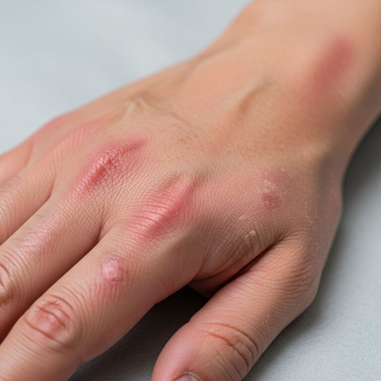
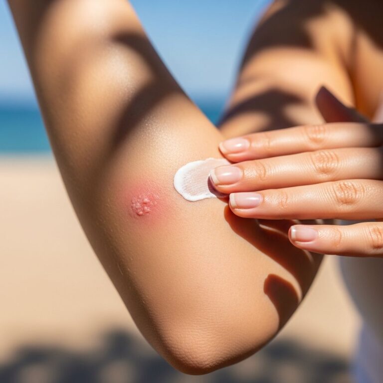
- Piloleiomyomas: Firm, skin-coloured to red-brown papules/nodules (2-20 mm), often multiple in clusters on trunk/extremities. Pain is hallmark: sharp, burning, triggered by cold, pressure, or stress. Lesions may urticate.
- Angioleiomyomas: Solitary, painful subcutaneous nodules (usually <1 cm) on legs, tender to pressure. Less cold-sensitive.
- Genital leiomyomas: Small, firm nodules on labia or scrotum; may cause dyspareunia.
In HLRCC, patients develop multiple cutaneous leiomyomas (often segmental) ± early-onset uterine fibroids and renal cancer risk.
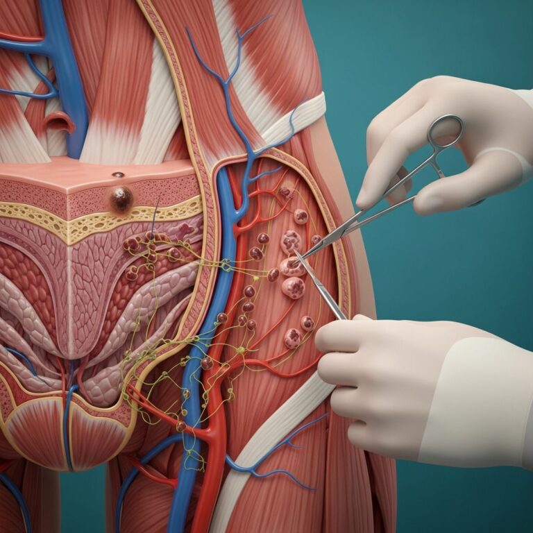
Diagnosis of leiomyoma
Diagnosis combines clinical suspicion and histopathology:
| Type | Key Diagnostic Features |
|---|---|
| Uterine | Ultrasound/MRI showing well-circumscribed masses; biopsy rarely needed unless atypical. |
| Cutaneous (Piloleiomyoma) | Skin biopsy: dermal spindle cells in fascicles, cigar-shaped nuclei, no atypia/mitoses. IHC: SMA+, desmin+. |
| Angioleiomyoma | Biopsy shows smooth muscle encircling vascular channels; SMA+. |
Differential includes dermatofibroma (dimple sign, less painful), neurofibroma (buttonhole sign), schwannoma, eccrine spiradenoma, and angiosarcoma (malignant). Pain history and biopsy distinguish. For multiple lesions, screen for HLRCC via FH gene testing and renal imaging.
How is leiomyoma treated?
Treatment targets symptoms, as tumours are benign:
- Solitary lesions: Surgical excision curative, low recurrence.
- Multiple cutaneous: High recurrence (50%); medical options first:
- Calcium channel blockers (nifedipine 30-60 mg/day) relieve pain via vasodilation.
- Alpha-blockers (phenoxybenzamine), antiepileptics (gabapentin), beta-blockers (propranolol), or antidepressants (imipramine).
- Botulinum toxin, cryotherapy, or laser ablation for refractory cases.
- Uterine: Watchful waiting, hormonal therapy (GnRH agonists), myomectomy, uterine artery embolization, or hysterectomy.
- HLRCC: Annual renal MRI; excision of symptomatic skin lesions.
Complications of leiomyoma
Most are uncomplicated, but:
- Pain impacting quality of life in cutaneous forms.
- Uterine: Anaemia, infertility, pregnancy loss, or sarcomatous change (<1%).
- HLRCC: Aggressive renal cell carcinoma (type 2 papillary), collecting duct carcinoma.
Prevention of leiomyoma
No proven prevention. For uterine: Maintain healthy weight, multiparity may reduce risk. Genetic counselling for HLRCC families.
Other conditions associated with leiomyoma
- Reed syndrome/HLRCC: FH mutations; cutaneous/uterine leiomyomas + renal cancer (15-30% lifetime risk).
- Rare: Gardner syndrome, HIV, CLL, polycythaemia.
Frequently Asked Questions
Are leiomyomas cancerous?
No, leiomyomas are benign. Malignant transformation to leiomyosarcoma is exceedingly rare (<0.5% in skin).
Why do cutaneous leiomyomas hurt?
Pain arises from smooth muscle contraction, amplified by cold/pressure on arrector pili or vessels.
Can leiomyomas be removed?
Yes, excision works for solitary lesions. Multiple require symptom management due to recurrence risk.
Should I worry about kidney cancer with skin leiomyomas?
If multiple/familial, screen for HLRCC with FH testing and renal imaging.
What is the best pain relief for piloleiomyoma?
Nifedipine is first-line; alternatives include gabapentin or surgery for isolated nodules.
References
- Multiple Cutaneous Leiomyomas of the Shoulder — PubMed Central. 2024. https://pmc.ncbi.nlm.nih.gov/articles/PMC12066081/
- Hereditary leiomyomatosis and renal cell cancer syndrome — DermNet NZ. 2023-10-01. https://dermnetnz.org/topics/hereditary-leiomyomatosis-and-renal-cell-cancer-syndrome
- Leiomyoma — DermNet NZ. 2023-10-01. https://dermnetnz.org/topics/leiomyoma
- Leiomyosarcoma pathology — DermNet NZ. 2023-10-01. https://dermnetnz.org/topics/leiomyosarcoma-pathology
- Angioleiomyoma pathology — DermNet NZ. 2023-10-01. https://dermnetnz.org/topics/angioleiomyoma-pathology
Similar Articles


Read full bio of Sneha Tete