Leishmaniasis: Causes, Symptoms, Diagnosis & Treatment
Comprehensive guide to leishmaniasis: Understanding parasitic infection, clinical forms, and treatment options.

Understanding Leishmaniasis: A Comprehensive Overview
Leishmaniasis is an infection caused by parasites from the genus Leishmania, transmitted to humans primarily through the bites of infected sandflies. This parasitic disease represents a significant global health concern, affecting millions of people in tropical and subtropical regions worldwide. The infection can manifest in multiple clinical forms, ranging from mild localized skin lesions to severe, life-threatening systemic disease. Understanding the nature of this infection, its transmission, clinical presentations, and available treatment options is essential for healthcare providers and individuals at risk.
What Is Leishmaniasis?
Leishmaniasis encompasses a group of diseases caused by infection with parasites from over 20 Leishmania species. These protozoan parasites are transmitted to humans through the bites of infected female sandflies, which are responsible for injecting the parasites into the bloodstream. Once inside the human body, these parasites invade macrophages—immune cells responsible for engulfing foreign organisms—and establish chronic infections that can persist for months or even years without treatment. The disease manifests differently depending on the Leishmania species involved and the individual’s immune response.
How Is Leishmaniasis Transmitted?
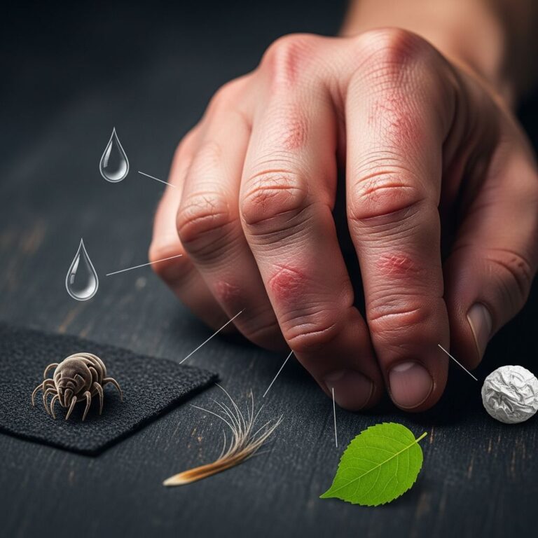
The transmission of leishmaniasis occurs exclusively through sandfly vectors. Over 90 sandfly species are known to transmit Leishmania parasites to humans. Female sandflies, which are active primarily during nighttime hours, require blood meals for reproduction. When an infected sandfly bites a human, it injects promastigotes—the infectious form of the parasite—into the skin. These promastigotes are rapidly phagocytosed by macrophages, where they transform into amastigotes and begin multiplying. The parasites can then spread to various tissues depending on the species and host factors.
Geographic distribution of leishmaniasis is closely tied to sandfly habitat and prevalence. The disease occurs throughout tropical and subtropical regions, with particular concentration in the Mediterranean area, Africa, Asia, and Latin America. High-risk areas include parts of South Asia, East Africa, and Brazil, where environmental conditions favor sandfly breeding and proliferation.
Types of Leishmaniasis
Leishmaniasis presents in three main clinical forms, each with distinct characteristics, severity levels, and treatment approaches. Understanding these forms is crucial for proper diagnosis and management.
Cutaneous Leishmaniasis (CL)
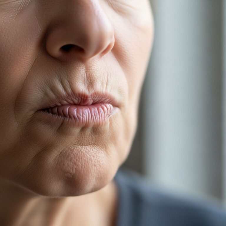
Cutaneous leishmaniasis is the most common form of the disease, affecting the skin at the site of sandfly inoculation. This localized form typically presents as skin lesions that develop weeks to months after infection. The lesions usually begin as small bumps or papules and progressively evolve into nodular plaques, eventually developing into characteristic ulcers with raised borders and central depressions. These ulcers may be covered by scabs or crusts and can persist for extended periods. Unless otherwise specified, cutaneous leishmaniasis refers to the localized form; however, much rarer variants include diffuse cutaneous leishmaniasis and disseminated cutaneous leishmaniasis, which can affect multiple areas simultaneously or recur repeatedly even with treatment.
The clinical course of cutaneous leishmaniasis involves progressive tissue destruction. Lesions typically are painless but may become painful if secondary bacterial infections occur or if they develop near joints. The healing process usually results in permanent atrophic scarring, which can cause cosmetic concerns and functional impairment. Even patients with single lesions commonly develop satellite lesions, regional lymphadenopathy, and nodular lymphangitis, creating a more widespread presentation than initially apparent.
Mucosal Leishmaniasis (ML)
Mucosal leishmaniasis typically develops as a progression from inadequately treated cutaneous disease, though it can occasionally present concomitantly with active skin lesions. This form usually becomes clinically evident within several years of the original cutaneous infection, sometimes developing decades later. The disease characteristically begins with persistent, unusual nasal symptoms including stuffiness or epistaxis, though oral or pharyngeal symptoms may also occur initially. Without treatment, mucosal leishmaniasis progresses to ulcerative destruction of the naso-oropharyngeal mucosa, potentially causing perforation of the nasal septum. This progression can become life-threatening if the larynx becomes involved, as infection can lead to airway obstruction.
Visceral Leishmaniasis (VL) or Kala-Azar
Visceral leishmaniasis, commonly known as kala-azar (meaning “black fever” in Hindi), represents the most severe form of the disease. This systemic infection affects internal organs, particularly the spleen, liver, and bone marrow. The disease is usually caused by Leishmania donovani and Leishmania infantum in the Eastern Hemisphere, and Leishmania infantum chagasi in the Western Hemisphere. While the incubation period generally ranges from weeks to months, asymptomatic infection can become clinically manifest years to decades after exposure in immunocompromised individuals. An estimated 50,000 to 90,000 new cases of visceral leishmaniasis occur worldwide annually, with only 25-45% reported to the World Health Organization.
Clinical Manifestations and Symptoms
Cutaneous Leishmaniasis Symptoms
Symptoms of cutaneous leishmaniasis typically begin a few weeks to several months after sandfly exposure. Initial presentation involves the development of skin lesions at bite sites, which progress through characteristic stages:
- Small bumps or papules that gradually enlarge
- Progression to nodular plaques with raised borders
- Development of central ulceration with distinctive “punched-out” appearance
- Formation of scabs or crusts covering the lesion base
- Associated regional lymphadenopathy and satellite lesions
- Painless lesions unless secondarily infected or irritated
In some cases, skin lesions may persist for months or years before spontaneous healing occurs. The healing process typically results in permanent scarring that may cause cosmetic disfigurement or functional impairment depending on lesion location.
Visceral Leishmaniasis Symptoms
Visceral leishmaniasis presents with systemic manifestations reflecting multi-organ involvement. Typical clinical features include:
- Irregular bouts of fever, often prolonged and recurrent
- Significant weight loss and cachexia (wasting)
- Hepatosplenomegaly with spleen enlargement typically more prominent than liver enlargement
- Pancytopenia including anemia, leukopenia, and thrombocytopenia
- Elevated total protein levels with low albumin and hypergammaglobulinemia
- Anemia causing paleness, dizziness, and shortness of breath
- Easy bruising and bleeding from reduced platelets
- Abdominal discomfort from organ enlargement
The severity and progression of visceral leishmaniasis varies considerably. In severe cases, immune system suppression becomes profound, rendering patients vulnerable to opportunistic infections including pneumonia and tuberculosis. Without prompt treatment, visceral leishmaniasis is fatal in over 95% of cases.
Diagnosis of Leishmaniasis
Accurate diagnosis of leishmaniasis requires clinical suspicion combined with laboratory confirmation. Healthcare providers should maintain awareness of leishmaniasis in patients presenting with appropriate clinical features and relevant exposure history, particularly travel to endemic areas.
Diagnostic approaches vary by leishmaniasis form. For cutaneous disease, diagnosis typically involves visualization of parasites through microscopy of lesion aspirates or biopsies stained with appropriate dyes. Culture techniques can isolate the parasite, allowing species identification and antimicrobial susceptibility testing. Serological tests and polymerase chain reaction (PCR) assays provide additional diagnostic modalities with varying sensitivity and specificity depending on disease stage and immune status.
Visceral leishmaniasis diagnosis frequently utilizes bone marrow or splenic aspiration for parasite visualization and culture. Serological testing demonstrates high sensitivity in immunocompetent individuals but may be less reliable in immunocompromised patients. PCR-based diagnostics offer enhanced sensitivity for detecting parasitic DNA in various tissue samples.
Treatment Options
Treatment of leishmaniasis depends on disease form, severity, geographic location, and individual patient factors including immune status. Therapeutic decisions require careful consideration of efficacy, toxicity profiles, and resistance patterns specific to the region and parasite species involved.
Cutaneous leishmaniasis treatment ranges from observation for self-limiting lesions to systemic therapy for extensive or problematic disease. Localized approaches include intralesional antimony injections or topical agents. Systemic therapy typically involves parenteral antimony compounds or alternative agents such as miltefosine or amphotericin B formulations. Mucosal leishmaniasis almost universally requires systemic therapy due to high rates of spontaneous progression and severe morbidity.
Visceral leishmaniasis mandates systemic treatment in nearly all cases. First-line therapies vary geographically, with pentavalent antimony compounds remaining standard in many regions, while amphotericin B formulations—particularly liposomal amphotericin B—are preferred in others due to superior efficacy and reduced toxicity. Miltefosine and paromomycin represent additional options with varying availability and efficacy across different geographic regions and patient populations.
Visceral and mucosal leishmaniasis become progressively more difficult to cure the longer they remain untreated. Treatment challenges intensify significantly in immunocompromised individuals, including those with HIV/AIDS, requiring adjusted therapeutic approaches and longer treatment courses. Relapse rates in immunocompromised patients remain concerning despite appropriate therapy, necessitating careful monitoring and consideration of maintenance therapy.
Complications of Leishmaniasis
Complications vary by disease form and severity. Cutaneous leishmaniasis complications primarily involve permanent scarring and potential disability depending on lesion location and number. Disseminated disease, though rare, can spread to multiple body sites.
Visceral and mucosal leishmaniasis complications are more serious and potentially life-threatening. Advanced visceral disease results in profound immune suppression, predisposing patients to severe opportunistic infections. Severe anemia from bone marrow involvement can become life-threatening without transfusion support. Mucosal disease can progress to catastrophic airway obstruction if laryngeal involvement occurs. Secondary infections are common in all forms, particularly in tropical environments with limited access to wound care.
Prevention and Risk Factors
Prevention of leishmaniasis focuses on reducing sandfly exposure. High-risk populations include travelers to endemic regions, particularly those engaging in outdoor activities during evening and nighttime hours when sandflies are active. Individuals with compromised immune systems, including those with HIV/AIDS, face substantially elevated risk for severe disease and reactivation of latent infection.
Prevention measures include appropriate use of insect repellents containing DEET on exposed skin, wearing protective clothing with long sleeves and pants, utilizing bed nets in endemic areas, and avoiding outdoor exposure during sandfly peak activity hours. Residents in endemic areas should implement environmental controls reducing sandfly habitats.
Frequently Asked Questions
Q: How long does it take for leishmaniasis symptoms to appear?
A: Symptoms typically appear within weeks to months of sandfly exposure, though cutaneous lesions occasionally first appear years later, particularly following trauma or immunosuppression. Visceral leishmaniasis may take months to years to become clinically evident, and asymptomatic infection can persist for decades before manifestation in immunocompromised individuals.
Q: Is leishmaniasis curable?
A: Yes, leishmaniasis is treatable with appropriate antiparasitic medications. Cutaneous disease often responds well to therapy, though treatment duration varies. Visceral and mucosal forms require aggressive systemic treatment and may be more challenging to cure, especially in immunocompromised patients. Early treatment significantly improves outcomes and reduces complications.
Q: Can leishmaniasis be transmitted person-to-person?
A: No, leishmaniasis cannot be transmitted directly between humans except in rare instances of blood transfusion or organ transplantation. Transmission occurs exclusively through sandfly vectors. However, pregnant women with visceral leishmaniasis can transmit infection to the fetus, making prenatal detection and treatment important.
Q: What should travelers do to prevent leishmaniasis?
A: Travelers to endemic areas should use insect repellent containing DEET on exposed skin, wear protective clothing covering arms and legs, utilize bed nets, and avoid outdoor exposure during peak sandfly activity hours (dusk to dawn). Seeking accommodation with screened windows or air conditioning provides additional protection.
Q: Are there vaccines available for leishmaniasis?
A: Currently, no widely available vaccines prevent leishmaniasis, though vaccine development research is ongoing. Prevention relies on reducing sandfly exposure through protective measures and environmental controls in endemic areas.
References
- Clinical Overview of Leishmaniasis — Centers for Disease Control and Prevention (CDC). 2024. https://www.cdc.gov/leishmaniasis/hcp/clinical-overview/index.html
- Leishmaniasis — World Health Organization (WHO). 2024. https://www.who.int/news-room/fact-sheets/detail/leishmaniasis
- Leishmaniasis: Causes, Symptoms, Diagnosis & Treatment — Cleveland Clinic. 2024. https://my.clevelandclinic.org/health/diseases/24539-leishmaniasis
- Cutaneous leishmaniasis — Britannica. 2024. https://www.britannica.com/science/cutaneous-leishmaniasis
- Visceral Leishmaniasis: Symptoms & Treatment — Rupa Health. 2024. https://www.rupahealth.com/post/visceral-leishmaniasis-symptoms-treatment
Similar Articles


Read full bio of medha deb