Lentigo: Causes, Features, and Treatment
Understanding lentigines: from causes and clinical features to effective treatment options.

What Is a Lentigo?
A lentigo is a pigmented flat or slightly raised lesion with a clearly defined edge that develops on the skin. Unlike an ephelis (freckle), a lentigo does not fade during winter months and persists year-round. The term “lentigo” is the singular form, while “lentigines” refers to multiple lesions of this type. These common skin lesions are characterized by localized areas of increased melanin production and are generally benign, though certain types warrant clinical attention.
Understanding Lentigines: Definition and Classification
Lentigines have been classified into several different types depending on their appearance, location on the body, causative factors, and whether they are associated with systemic diseases or conditions. The most common classifications include solar lentigines, which result from ultraviolet radiation exposure, and syndromic lentigines, which are associated with inherited genetic conditions. This classification system helps clinicians distinguish between benign age-related lesions and those that may indicate underlying systemic pathology.
Types of Lentigo
Solar Lentigo (Age Spots)
Solar lentigo, also known as senile lentigo, age spot, or liver spot, is a benign pigmented macule that appears on chronically sun-exposed skin. This type is the most common form of lentigo and develops when ultraviolet (UV) radiation causes pigmented cells called melanocytes in the skin to multiply. Solar lentigines typically appear on sun-exposed areas of the body, including the face, hands, shoulders, and arms, and are particularly common in people over age 40, though younger individuals can develop them as well. The spots may grow slowly over many years or appear suddenly, and they may have rounded or uneven edges.
Lentigo Maligna
Lentigo maligna is a slow-growing subtype of melanoma in situ that commonly develops on chronically sun-damaged skin, particularly in older adults. It most frequently affects the head and neck region and presents as an irregular brown macule or patch on chronically sun-damaged skin. On visual inspection, lesions may appear light-brown to black, display color variegation, appear asymmetric, and tend to have an ill-defined border. Unlike benign solar lentigines, lentigo maligna poses unique diagnostic challenges due to its ability to clinically and histopathologically resemble benign pigmented lesions, such as solar lentigines or seborrheic keratoses. As lesions enlarge, they may develop skip areas with a patchy, noncontiguous pattern. Advanced tumors may produce pain, burning, itching, or bleeding.
PUVA-Induced Lentigo
A variant known as psoralen ultraviolet A (PUVA)-induced lentigo is seen in approximately 50% of patients with at least 6 years of PUVA therapy. This type may sometimes be distinguished by large, somewhat atypical melanocytes on histopathology and can appear on any body surface exposed to PUVA treatment, including the genitalia.
Ink Spot Lentigo
An ink spot (sunburn) lentigo is another variant that appears as small black or dark gray macules resembling ink spots with reticulated borders, often located on the shoulders.
Causes of Lentigo
Ultraviolet Radiation Exposure
Common forms of lentigo are due to exposure to ultraviolet radiation, which is the primary cause of solar lentigines. UV exposure triggers melanocyte proliferation and increased melanin production, leading to the characteristic pigmented lesions seen in individuals with significant sun exposure history.
Ionising Radiation
Ionising radiation, such as that from radiation therapy, can also cause lentigines. This represents an important consideration in patients who have undergone therapeutic radiation for malignancy or other conditions.
Genetic and Syndromic Causes
Several familial syndromes are associated with widespread lentigines, and these conditions originate from mutations in Ras-MAP kinase, mTOR signalling, and PTEN pathways. Genetic syndromes associated with lentigines include:
- Bannayan-Riley-Ruvalcaba syndrome: This condition causes a larger-than-normal head, noncancerous tumors, and dark spots on the genitals.
- Cowden syndrome: This disorder causes many noncancerous growths called hamartomas to form on the body.
- Noonan syndrome: This condition causes lentigines to form on many different parts of the body.
- Peutz-Jeghers syndrome: This condition causes noncancerous growths to form in the stomach and intestines, with a higher risk of developing cancer in the lifetime. Children with Peutz-Jeghers often develop small dark spots on their faces, as well as spots on their lips, hands, feet, genitals, and inside the mouth. It is common for these spots to fade with age.
Clinical Features and Presentation
Lentigines typically present as pigmented lesions with distinct clinical characteristics. Unlike freckles, which fade seasonally, lentigines maintain their pigmentation throughout the year. The increase in melanin is often associated with specific dermal changes and represents a localized area of melanocytic hyperactivity. Most lentigines are relatively straightforward to diagnose clinically, though certain cases may present diagnostic challenges, particularly when distinguishing lentigo maligna from benign solar lentigines.
Appearance and Characteristics
- Flat or slightly raised lesions with clearly defined edges
- Light-brown to black coloration, depending on type and individual factors
- Potential color variegation within individual lesions
- Asymmetric appearance in some cases
- Variable sizes, ranging from small macules to larger patches
- Typically asymptomatic, though advanced lesions may cause discomfort
- Persistent pigmentation regardless of season
Histopathology
Histopathologic examination of lentigo reveals characteristic findings that aid in diagnosis. In solar lentigines, the lesions show increased melanin production and melanocyte activity. Lentigo maligna, however, is characterized by a proliferation of atypical melanocytes along the dermal-epidermal junction. Early lentigo maligna may be challenging to distinguish from benign changes of chronic solar damage, which also induce melanocytic hyperplasia.
In lentigo maligna, melanocytes are typically singly arranged, although nests and multinucleated cells may be seen. The tumor cells often have a conspicuous cytoplasmic retraction artifact, and nuclei are often enlarged, hyperchromatic, and angulated. Background changes of chronic sun damage are usually present, including solar elastosis, epidermal atrophy, and effacement of the rete ridges. Pigmentation levels vary but may be abundant, and melanophages often populate the papillary dermis.
Lymphocytes typically localize to the superficial dermis; however, marked lymphocytic infiltration may indicate tumor invasion and warrants closer histopathologic evaluation. For this reason, a control biopsy of non-lesional but sun-damaged skin is sometimes recommended to aid differentiation.
Distinction from Other Skin Lesions
Although lentigo spots can resemble certain types of skin cancer, most lentigines, particularly solar lentigines, are noncancerous. However, lentigo maligna represents a significant exception, as it is classified as melanoma in situ and carries the potential for malignant progression. The key distinguishing feature between a lentigo and a freckle (ephelis) is that lentigines maintain their pigmentation throughout the year, whereas freckles fade during winter months when sun exposure decreases.
When Lentigo Becomes Lentigo Maligna Melanoma
By definition, lentigo maligna remains confined to the epidermis. Once the lesion becomes invasive—meaning atypical melanocytes extend into the dermis—the diagnosis changes to lentigo maligna melanoma (LMM), which carries a significantly worse prognosis. As lentigo maligna becomes more invasive and progresses to LMM, junctional nests become larger and more spindled. Invasion is typically superficial, consisting of isolated or small aggregates of spindled cells in the papillary dermis. Desmoplasia and perineural invasion may occur, and the desmoplastic component may be mistaken for a scar.
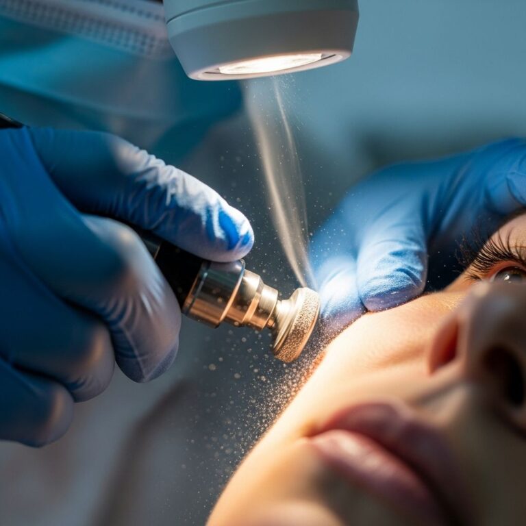
Treatment Options
Assessment and Monitoring
Although lentigo spots are often caused by sun damage to your skin, benign solar lentigines are not cancerous or harmful and do not require medical treatment. Some people choose to have lentigines removed because they dislike how they look on their skin, which represents a cosmetic choice rather than a medical necessity. However, careful evaluation is essential to distinguish benign solar lentigines from lentigo maligna.
Surgical Approaches
Surgical excision is the treatment of choice for lentigo maligna, with Mohs micrographic surgery (MMS) emerging as a surgical option that may prove superior to wide local excision in certain cases. These surgical approaches aim to balance oncologic control with cosmetic and functional outcomes, particularly in cosmetically sensitive areas such as the face.
Non-Surgical Removal Methods
A lentigo can grow very slowly over many years, or it can appear suddenly. Some types of lentigo can disappear on their own over time, but most do not go away without intervention. Other types can only be removed with treatment. For individuals seeking cosmetic removal of benign solar lentigines, various non-surgical modalities may be considered, though these are typically pursued for aesthetic rather than medical reasons.
Prevention Strategies
Since UV radiation exposure is the primary cause of solar lentigines, sun protection measures are essential for prevention. Individuals should employ comprehensive sun protection strategies, including:
- Regular use of broad-spectrum sunscreen with appropriate sun protection factor (SPF)
- Protective clothing, including wide-brimmed hats and long sleeves
- Avoiding peak sun exposure hours (typically 10 AM to 4 PM)
- Seeking shade whenever possible during high UV index periods
- Avoiding prolonged or intense sun exposure, particularly for individuals at higher risk
Clinical Considerations
Lentigo maligna and LMM present diagnostic and treatment challenges due to their clinical mimicry of benign lesions, their occurrence on background sun-damaged skin (which confounds histopathologic differentiation between true tumor and benign melanocytic activation), and their occurrence on the head and neck—a cosmetically and functionally sensitive area. Thus, maintaining clinical diligence and having a high index of suspicion are key to early diagnosis, ensuring optimal treatment outcomes, and minimizing patient morbidity and mortality.
An in-depth knowledge of the distinguishing features and distinctive therapeutic approaches of lentigo maligna and LMM is essential to ensure accurate diagnosis, optimal treatment planning, and improved patient outcomes.
Frequently Asked Questions
Q: Are lentigines dangerous or cancerous?
A: Most lentigines, particularly solar lentigines, are benign and noncancerous. However, lentigo maligna is a type of melanoma in situ and requires medical evaluation and treatment to prevent progression to invasive melanoma.
Q: Why don’t lentigines fade in winter like freckles do?
A: Unlike freckles (ephelides), lentigines maintain their pigmentation throughout the year because they represent a more permanent increase in melanin production and melanocyte activity in the skin.
Q: Can lentigines be removed?
A: Some types of lentigo can disappear on their own over time, but most do not go away without intervention. For cosmetic removal of benign solar lentigines, various treatment options may be available, though removal is typically pursued for aesthetic rather than medical reasons.
Q: What should I do if I notice a new pigmented lesion or changes in an existing one?
A: You should see a dermatologist if you notice a new skin growth, a change in an existing mole, or any skin changes that seem unusual. Early evaluation is particularly important to distinguish between benign solar lentigines and potentially malignant lentigo maligna.
Q: Are there genetic syndromes associated with lentigines?
A: Yes, several familial syndromes are associated with widespread lentigines, including Bannayan-Riley-Ruvalcaba syndrome, Cowden syndrome, Noonan syndrome, and Peutz-Jeghers syndrome. These conditions originate from mutations in various genetic pathways.
References
- Lentigo Maligna Melanoma — National Institutes of Health, National Center for Biotechnology Information. 2024. https://www.ncbi.nlm.nih.gov/books/NBK482163/
- Lentigo (Liver Spots): Pictures, Causes, Removal & Prevention — Healthline Media. 2025. https://www.healthline.com/health/lentigo
- Solar Lentigo — VisualDx. 2025. https://www.visualdx.com/visualdx/diagnosis/solar+lentigo
- Lentigo: Causes, Features, and Treatment — DermNet. 2024. https://dermnetnz.org/topics/lentigo
- Lentigo (syn. lentigine) — Primary Care Dermatology Society. 2023. https://www.pcds.org.uk/clinical-guidance/lentigo
- Lentigo (Age Spots) — Glick Skin Institute. 2024. https://glickskin.com/services/lentigo-age-spots/
Similar Articles


Read full bio of Sneha Tete