Lepra Reactions: 3 Types, Diagnosis And Treatment
Understanding the inflammatory complications of leprosy: Type 1 and Type 2 reactions, clinical features, diagnosis, and management strategies.

Lepra reactions, also known as leprosy reactions, are acute inflammatory episodes that complicate the course of leprosy, a chronic infection caused primarily by Mycobacterium leprae. These reactions arise from immunological responses, including T-cell mediated hypersensitivity in Type 1 reactions and immune complex vasculitis in Type 2 reactions. They can occur before, during, or after multidrug therapy (MDT) and pose significant risks for nerve damage, deformity, and systemic complications if not managed promptly.
Understanding lepra reactions is crucial for clinicians, as they affect up to 50% of leprosy patients and are a leading cause of disability. This article details their classification, clinical presentation, histopathology, risk factors, diagnosis, and treatment, drawing from authoritative sources like WHO guidelines and peer-reviewed studies.
What are lepra reactions?
Lepra reactions represent hypersensitivity responses to mycobacterial antigens in leprosy patients. They are categorized into three main types: Type 1 (reversal reaction), Type 2 (erythema nodosum leprosum or ENL), and rarely, Lucio phenomenon. Type 1 reactions involve a cell-mediated (Type IV) immune response, while Type 2 results from immune complex deposition causing vasculitis.
These episodes can manifest suddenly, exacerbating skin lesions and nerve involvement. In multibacillary (MB) leprosy, particularly lepromatous forms, reactions are more frequent due to high bacillary loads. Male patients at the lepromatous spectrum end show higher incidence. Reactions may persist for months if untreated, leading to irreversible nerve function loss and deformities.
Who gets lepra reactions?
Lepra reactions primarily affect individuals with borderline or lepromatous leprosy. Type 1 reactions occur in borderline tuberculoid (BT), borderline borderline (BB), and borderline lepromatous (BL) cases. Type 2 reactions are seen in lepromatous leprosy (LL) or BL patients with high bacillary indices.
- Patients starting MDT, especially within the first months.
- Postpartum women due to immune shifts during puerperium.
- Individuals with heavy skin infiltration or BI ≥3, more common in males with MB leprosy.
- Up to 60% of MB patients experience reactions, with Type II less frequent but more severe.
Risk factors include high bacterial load, skin infiltration, and nerve involvement at diagnosis. Disability is more prevalent in Type 2 (61.9%) than Type 1 (36.4%).
Type 1 lepra reaction
Type 1 lepra reaction (T1R), or reversal reaction, is a delayed-type hypersensitivity response affecting skin and peripheral nerves without systemic symptoms. It typically arises in borderline leprosy patients shortly after starting treatment.
Skin signs of type 1 lepra reaction
Skin manifestations include inflammation of existing plaques, which become erythematous, swollen, and warm. New lesions may appear, with edema, ulceration, or scaling.
- Erythematous, raised plaques.
- Nodule formation on lesions.
- Facial edema in severe cases.
Nerve involvement in type 1 lepra reaction
Nerve damage is hallmark, with tender, enlarged peripheral nerves adjacent to lesions. Symptoms include pain, paresthesia, weakness, and sensory loss, potentially leading to paralysis and deformity.
- Common nerves: ulnar, median, peroneal, posterior tibial.
- >2 nerves involved in 60-81% of cases.
Severity classification: Mild (few plaques, no nerve damage); Severe (multiple plaques, nerve impairment).
Clinical diagnosis of type 1 lepra reaction
Suspected in borderline leprosy patients developing symptoms post-treatment initiation. No routine labs show changes in neutrophils or CRP, but elevated serum chemokine 10 may occur. Skin biopsy reveals dermal edema, lymphocyte infiltration, and granulomas.
Management of type 1 lepra reaction
Mild cases need supportive care: rest, analgesia, wound care. Severe cases with neuritis require prompt steroids (prednisolone 1 mg/kg/day, tapered over 12 weeks) and nerve decompression if needed. Continue MDT uninterrupted.
Untreated, reactions persist months, risking permanent damage.
Type 2 lepra reaction
Type 2 lepra reaction (T2R), or ENL, is an immune complex-mediated vasculitis affecting skin and viscera in LL/BL patients. It features fever, malaise, and painful nodules.
Who gets type 2 lepra reaction?
Occurs in lepromatous or BL leprosy with high BI. Associated with skin infiltration, disability, and systemic features like uveitis or orchitis. Less frequent than Type 1 but more debilitating.
Clinical features of type 2 lepra reaction
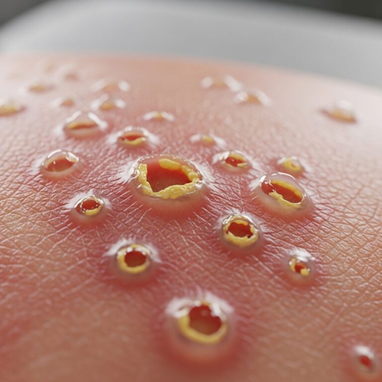
Skin: Tender erythematous nodules/papules on limbs/face, evolving to pustules, bullae, or necrosis. Extracutaneous: fever, arthralgia, lymphadenitis, neuritis, iridocyclitis.
- Multiple evanescent nodules (days-long).
- Pustular discharge with bacilli.
- Systemic: fatigue, high ESR/CRP.
Diagnosis of type 2 lepra reaction
Clinical suspicion in LL/BL with fever and nodules. Labs: leukocytosis, high CRP/ESR. Biopsy (<24h nodule): panniculitis, neutrophils, vasculitis, bacilli.
Treatment of type 2 lepra reaction
Mild: NSAIDs, analgesics, rest. Severe: prednisolone (1 mg/kg), thalidomide (100-400 mg/day for males/non-pregnant), or alternatives like clofazimine. Immunosuppressants (ciclosporin, methotrexate) for refractory cases.
Thalidomide is highly effective but contraindicated in pregnancy.
Lucio phenomenon
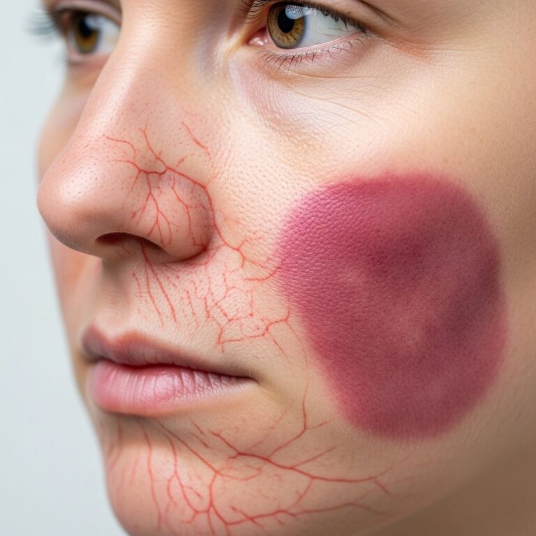
Rare necrotizing vasculitis in diffuse lepromatous leprosy (Lucio leprosy). Features irregular angular erythematous patches progressing to hemorrhagic infarcts and stellate scars. Biopsy shows endothelial proliferation and thrombosis without neutrophils. Treat with steroids/clofazimine.
Differential diagnosis
| Condition | Key Features | Differentiator |
|---|---|---|
| Drug eruption | Diffuse rash post-MDT | Biopsy lacks leprosy features |
| Cellulitis | Unilateral, bacterial | Culture positive, no nodules |
| Sweet syndrome | Fever, plaques | No bacilli, neutrophilic dermatosis |
| Polyarteritis nodosa | Deep nodules | No leprosy history |
Investigations
Standard: BI slit-skin smear, nerve exam. Specific: Biopsy for confirmation, inflammatory markers (ESR, CRP), CBC for leukocytosis.
Management
Continue MDT. Tailor to type/severity: steroids first-line, thalidomide for ENL. Monitor nerves with voluntary muscle testing/sensory charts. Prevent disability via physiotherapy.
Complications
- Nerve damage: anesthesia, weakness, ulcers.
- Deformities: claw hand, foot drop.
- Systemic: blindness (uveitis), infertility (orchitis).
Prevention
Early MDT, reaction monitoring in high-risk (MB, high BI). Prophylactic steroids/thalidomide in select cases.
Immunopathogenesis
T1R: Th1/Th17 cytokines (IFN-γ, TNF-α), CD8 T-cells damaging nerves. T2R: Immune complexes, complement activation, neutrophil influx.
Frequently Asked Questions
What causes lepra reactions?
Immunological responses to M. leprae antigens, triggered by MDT or immune shifts.
Can lepra reactions occur after MDT completion?
Yes, due to residual antigens; ongoing monitoring needed.
Is thalidomide safe for all ENL patients?
No, contraindicated in pregnancy; use alternatives like steroids.
How long do reactions last?
Weeks to months untreated; resolve faster with therapy.
Do reactions indicate treatment failure?
No, they signify immune activity; continue MDT.
References
- The Lepra Reaction With Necrotizing Skin Lesions: A Report of Six Cases — Moschella SL. JAMA Dermatology. 1967-06. https://jamanetwork.com/journals/jamadermatology/fullarticle/530270
- Leprosy Reactions: Frequency and Risk Factors — Herald Open Access. 2016. https://www.heraldopenaccess.us/openaccess/leprosy-reactions-frequency-and-risk-factors
- Lepra reactions — DermNet NZ. 2023. https://dermnetnz.org/topics/lepra-reactions
- Leprosy — Walker SL, et al. StatPearls [Internet]. NCBI Bookshelf. 2023-08-07. https://www.ncbi.nlm.nih.gov/books/NBK559307/
- Leprosy reactions: Unraveling immunological mechanisms — Bobosha K, et al. PMC. 2024. https://pmc.ncbi.nlm.nih.gov/articles/PMC11180982/
- Leprosy Reactions — International Textbook of Leprosy. 2023. https://www.internationaltextbookofleprosy.com/clinical-sciences/clinical-aspects/leprosy-reactions
Similar Articles


Read full bio of medha deb