Leukemia Cutis: Key Insights Into Diagnosis & Treatment
Understanding leukaemia cutis: skin manifestations of leukaemia, from symptoms and diagnosis to treatment options and prognosis.

Leukaemia cutis refers to the infiltration of the skin by neoplastic leucocytes, resulting in clinically characteristic skin lesions that may erupt at any time during the course of leukaemia. It signifies advanced disease in most cases and carries a poor prognosis.
What is leukaemia cutis?
Leukaemia cutis is a dermatological manifestation of leukaemia where malignant leucocytes from the peripheral blood infiltrate the skin, primarily the dermis. This infiltration leads to visible and palpable skin changes, ranging from small papules to large nodules and plaques. It is relatively uncommon, occurring in approximately 10-15% of leukaemia patients overall, but the incidence varies by leukaemia subtype. Acute myeloid leukaemia (AML) shows the highest association, with up to 20% of cases developing skin involvement, while it is rarer in chronic forms like chronic lymphocytic leukaemia (CLL), affecting only about 4%.
The exact mechanism driving leukaemia cells to infiltrate the skin remains unclear, though factors such as homing receptors on malignant cells and favourable skin microenvironment may play roles. In most instances (55-77%), skin lesions appear in patients already diagnosed with leukaemia due to abnormal blood counts or bone marrow findings. However, in 23-44% of cases, lesions coincide with the initial presentation of systemic leukaemia, and in 2-3%, they precede detectable blood or bone marrow involvement—a condition termed aleukaemic leukaemia cutis. This aleukaemic form eventually progresses to overt leukaemia, often AML.
Children with congenital leukaemia exhibit a higher rate, around 25-30%, sometimes presenting as ‘blueberry muffin syndrome’ with purpuric nodules due to dermal erythropoiesis.
Who gets leukaemia cutis?
Leukaemia cutis can affect any age but is more common in adults with acute leukaemias. It predominantly occurs in patients with myeloid lineage leukaemias, particularly:
- Acute myeloid leukaemia (AML): Most frequent, up to 20% incidence; associated with monocytic subtypes (FAB M4/M5).
- Acute lymphoblastic leukaemia (ALL): Less common, around 10-20% in children.
- Adult T-cell leukaemia/lymphoma (ATLL): Rare but highly associated when present.
- Chronic myeloid leukaemia (CML) and CLL: Rare, <5%; in CLL, it often signals progression or Richter’s transformation.
Risk factors include high leucocyte counts, monocytic differentiation, and extramedullary disease. Patients with relapsed or refractory leukaemia are also prone.
What causes leukaemia cutis?
The primary cause is the dissemination of neoplastic leucocytes from blood or bone marrow into the skin via haematogenous spread or local invasion. Leukaemia cells adhere to dermal endothelium using adhesion molecules and migrate into the tissue, proliferating there. Genetic mutations in leukaemia cells, such as those promoting tissue tropism, contribute. It is not triggered by external factors but reflects the aggressiveness of the underlying haematological malignancy. Secondary skin changes, like infections or drug reactions, can mimic or complicate leukaemia cutis in leukaemia patients.
What are the clinical features of leukaemia cutis?
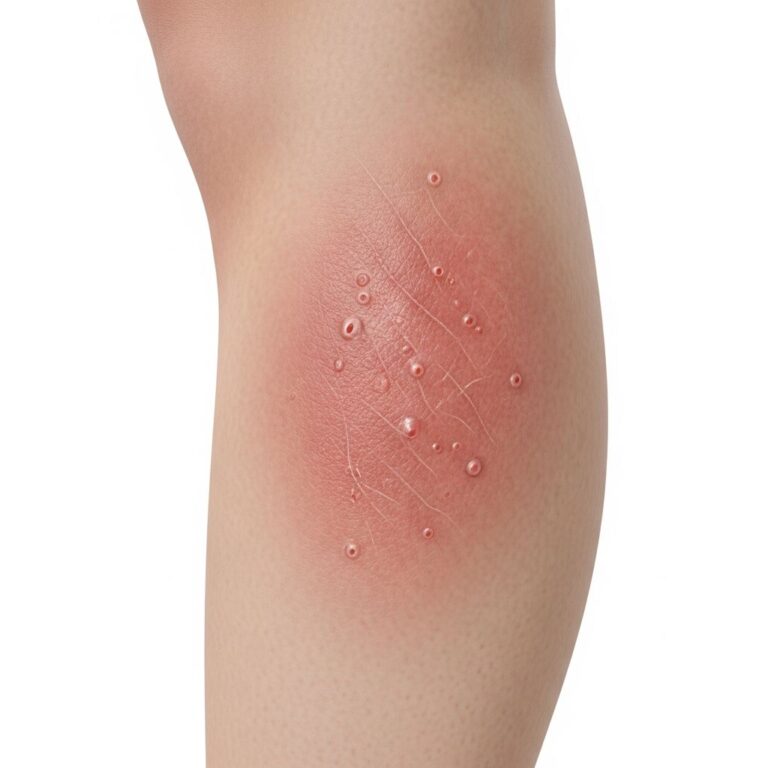
Skin lesions vary widely but commonly include:
- Papules and nodules (60%): Firm, rubbery, 5-10 mm; red, purple, skin-coloured, or violaceous.
- Plaques: Indurated, erythematous patches.
- Rare forms: Erythroderma, ulcers, blisters, bullae; Sweet syndrome-like pustules in AML.
Lesions often appear on the trunk, extremities, face, and scalp. They are usually asymptomatic but may itch, ulcerate, or bleed. Specific patterns include:
| Leukaemia Type | Typical Lesions |
|---|---|
| AML (monocytic) | Gum hypertrophy, oral ulcers, papules/nodules |
| ATLL | Nodules, plaques, erythroderma |
| CLL | |
| Congenital ALL | Blueberry muffin spots |
Systemic symptoms like fever suggest infection, common due to immunosuppression.
How is leukaemia cutis diagnosed?
Diagnosis combines clinical suspicion, haematological correlation, and histopathology:
- Clinical exam and history: Lesions in known leukaemia patient raise suspicion.
- Skin biopsy: Essential; shows neoplastic leucocytes in dermis/epidermis. Immunophenotyping confirms lineage (e.g., myeloperoxidase for myeloid).
- Blood/bone marrow tests: Peripheral smear, flow cytometry, cytogenetics.
- Differential diagnosis: Includes cutaneous lymphomas, drug eruptions, infections, vasculitis. Aleukaemic cases require vigilant monitoring.
What is the treatment for leukaemia cutis?
Treatment targets the underlying leukaemia, as local therapy alone fails due to systemic disease:
- Systemic chemotherapy: Induction regimens (e.g., 7+3 for AML); leads to lesion regression in 70-90% of responders.
- Targeted therapies/Immunotherapy: Tyrosine kinase inhibitors for CML, monoclonal antibodies for lymphoid leukaemias.
- Stem cell transplant: For high-risk/relapsed cases.
- Local therapies: Radiation (electron beam), surgery for symptomatic/palliative lesions; phototherapy rarely.
- Symptomatic care: Topical steroids, lidocaine, menthol for pain/pruritus; antimicrobials for infections; moisturizers (e.g., Biafine) for radiation burns.
Watchful waiting applies to indolent forms like early CLL.
What is the prognosis for leukaemia cutis?
Leukaemia cutis indicates advanced, high-risk disease with poor prognosis. Median survival post-diagnosis is 6-12 months, worse in AML (monocytic) and aleukaemic forms. It correlates with extramedullary spread and chemotherapy resistance. Early systemic therapy improves skin clearance but not overall survival significantly.
Related skin conditions in leukaemia patients
Leukaemia patients face multiple dermatoses:
- Infections: Bacterial (impetigo), fungal (candida), viral (HSV, VZV) due to neutropenia.
- Drug reactions: Morbilliform rashes, Stevens-Johnson from antibiotics/chemo.
- Neutrophilic dermatoses: Sweet syndrome, pyoderma gangrenosum (PG) in AML/myelodysplasia.
- Paraneoplastic: Itchy eruptions, acrokeratosis paraneoplastica.
- Pallor/petechiae/purpura: From anaemia/thrombocytopenia.
Frequently Asked Questions
Q: Is leukaemia cutis painful?
A: Most lesions are painless, but ulceration, infection, or secondary changes can cause discomfort.
Q: Can leukaemia cutis be the first sign of leukaemia?
A: Yes, in 2-7% of cases (aleukaemic leukaemia cutis), but bone marrow involvement follows.
Q: Does treating the skin cure leukaemia cutis?
A: No; systemic leukaemia therapy is required, as skin lesions recur if underlying disease persists.
Q: What does leukaemia cutis look like in children?
A: Often blueberry muffin rash—purplish nodules from dermal haematopoiesis.
Q: How common is leukaemia cutis in AML?
A: Up to 20%, especially monocytic subtypes; portends worse prognosis.
References
- Leukemia cutis: Symptoms, pictures, treatment, and outlook — Medical News Today. 2023-05-15. https://www.medicalnewstoday.com/articles/leukemia-cutis
- Leukemia Cutis: Photos, Prevalence, and Treatment — MyLeukemiaTeam. 2024-02-10. https://www.myleukemiateam.com/resources/leukemia-cutis-an-overview
- Leukemia Cutis – StatPearls — NCBI Bookshelf. 2023-07-17. https://www.ncbi.nlm.nih.gov/books/NBK541136/
- What Leukemia Does to Your Skin — Healthgrades. 2024-01-22. https://resources.healthgrades.com/right-care/leukemia/what-leukemia-does-to-your-skin
- Leukaemia Cutis — Leukaemia Care. 2023-11-05. https://www.leukaemiacare.org.uk/support-and-information/latest-from-leukaemia-care/blog/leukaemia-cutis/
- Aleukemic leukemia cutis — Cleveland Clinic Journal of Medicine. 2019-02-01. https://www.ccjm.org/content/86/2/85
- Leukaemia cutis – DermNet — DermNet NZ. 2024-06-12. https://dermnetnz.org/topics/leukaemia-cutis
Similar Articles


Read full bio of Sneha Tete