Managing Leukocoria: Treatment Options and Outcomes
Comprehensive guide to diagnosing and treating white pupil conditions in children and adults.

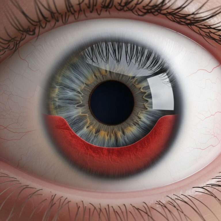
Leukocoria, commonly referred to as the “white pupil” or “cat’s eye pupil,” represents a significant clinical finding that warrants immediate medical evaluation. This abnormal reflection occurs when light strikes the pupil and produces a white glow instead of the typical red reflex seen in healthy eyes. The presence of leukocoria can indicate numerous underlying eye conditions ranging from benign to life-threatening, making prompt diagnosis and appropriate treatment essential for preserving vision and overall eye health.
Understanding the Diagnostic Foundation
Before any treatment plan can be established, a comprehensive ophthalmologic evaluation must be performed to identify the specific condition causing the white pupil appearance. The diagnostic process begins with a detailed patient history, including the age of onset, whether the condition affects one or both eyes, and any accompanying symptoms such as vision changes, eye redness, or behavioral changes in children.
The red reflex examination represents the cornerstone of initial assessment. During this examination, an ophthalmologist uses a light source to evaluate the transparency of the eye’s optical media and the health of the retina. Any abnormality in this reflex—particularly a white or absent reflex—triggers further investigation.
Advanced imaging techniques provide crucial information for accurate diagnosis:
- Dilated eye examination: Pupillary dilation allows clearer visualization of posterior segment structures and lesions.
- Ultrasound imaging: B-scan ultrasonography helps evaluate retinal structures when media opacities obscure direct visualization.
- Magnetic resonance imaging (MRI): This imaging modality is particularly valuable for evaluating retinoblastoma and determining whether the tumor has spread beyond the eye.
- Computed tomography (CT): CT scanning can assess bone involvement and extraocular extension in malignant conditions.
- Optical coherence tomography (OCT): This non-invasive technique provides detailed cross-sectional images of retinal structures.
Condition-Specific Treatment Approaches
The underlying cause of leukocoria determines the appropriate treatment strategy. Different conditions require distinctly different management approaches, ranging from observation to aggressive intervention.
Management of Retinoblastoma
Retinoblastoma, a malignant tumor of the retina, represents one of the most serious causes of leukocoria and is the most common intraocular malignancy in children. Treatment decisions depend on multiple factors including tumor size, location, laterality, and whether metastatic spread has occurred.
Contemporary retinoblastoma management includes several evidence-based approaches:
- Chemotherapy: Systemic chemotherapy may be administered intravenously, directly into the blood vessels supplying the eye (intra-arterial chemotherapy), or injected directly into the vitreous cavity (intravitreal chemotherapy) to target malignant cells while preserving the globe and vision when possible.
- Radiation therapy: External beam radiation or brachytherapy (internal radiation) may be employed for tumors that do not respond adequately to chemotherapy alone.
- Cryotherapy: This freezing technique destroys tumor tissue and may be combined with other modalities for smaller lesions.
- Laser photocoagulation: Targeted laser energy destroys tumor cells in selected cases.
- Enucleation: Surgical removal of the affected eye becomes necessary when the tumor is advanced, vision cannot be salvaged, or other treatments have failed.
Addressing Congenital Cataracts
Congenital cataracts, which involve clouding of the lens present from birth, frequently cause leukocoria in newborns and young infants. The management approach depends on the density and location of the opacity and whether it significantly impairs vision development.
Treatment may include observation in cases of mild, peripheral cataracts that do not affect visual axis. However, dense cataracts or those affecting the central lens typically require surgical removal to prevent irreversible vision loss and amblyopia (lazy eye). Following cataract extraction, optical rehabilitation through glasses, contact lenses, or intraocular lens implants restores focusing ability and promotes normal visual development.
Treating Coats Disease
Coats disease, characterized by abnormal retinal blood vessels and exudative changes, presents with leukocoria and progressive retinal detachment if untreated. The progressive nature of this condition necessitates active intervention.
Treatment modalities include laser photocoagulation to ablate abnormal vascular areas and reduce exudation, cryotherapy to destroy ischemic retinal regions, anti-vascular endothelial growth factor (anti-VEGF) injections to reduce abnormal vessel formation, and corticosteroids to control inflammation. In advanced cases with extensive retinal detachment, vitrectomy may be required.
Managing Retinopathy of Prematurity
Retinopathy of prematurity (ROP) develops in premature infants whose retinal blood vessels failed to fully develop before birth. Early detection through systematic screening of at-risk infants allows timely intervention before severe vision loss occurs.
Treatment approaches vary by disease severity. Anti-VEGF injections and laser photocoagulation represent the primary interventions, with anti-VEGF therapy increasingly preferred for aggressive posterior disease. These treatments halt abnormal vessel proliferation and reduce the risk of retinal detachment. In cases where tractional retinal detachment develops despite initial treatment, vitrectomy may restore some vision.
Addressing Persistent Fetal Vasculature
Persistent fetal vasculature (PFV) occurs when embryonic blood vessels in the eye fail to regress normally, resulting in fibrovascular membranes that can distort ocular structures. Most cases are non-progressive and require only monitoring, but some develop complications requiring intervention.
Surgical intervention becomes necessary when PFV causes progressive lens opacity, retinal detachment, or intractable glaucoma. Vitrectomy and membrane peeling may restore some vision by removing obstructing fibrovascular tissue.
Treating Ocular Infections and Inflammation
Leukocoria caused by infectious processes such as ocular toxoplasmosis or parasitic infections requires pathogen-specific antimicrobial therapy. Toxoplasma gondii infection responds to combinations of anti-parasitic medications, while toxocariasis treatment involves similar anti-parasitic agents.
Inflammatory conditions causing leukocoria respond to anti-inflammatory therapy. Corticosteroids applied topically, injected locally, or administered systemically reduce inflammation of the uvea and associated tissues. Immunosuppressive agents may be required for chronic or refractory cases.
Managing Retinal Detachment
Retinal detachment, whether due to trauma or disease, requires urgent surgical intervention to reattach the retina and preserve remaining vision. Pneumatic retinopexy, scleral buckle surgery, or vitrectomy may be employed depending on detachment characteristics, with the goal of restoring retinal anatomy and halting progressive vision loss.
Age-Specific Considerations in Treatment Planning
Treatment decisions differ substantially between pediatric and adult populations. Young children require approaches that account for developing visual systems, compliance challenges, and the critical period for normal visual development. Developmental abnormalities in the visual pathways can result in permanent vision loss if treatment is delayed during this sensitive window.
Adults with leukocoria may tolerate treatment differently and may have established visual preferences and lifestyle considerations that influence management decisions. The presence of comorbidities in older adults also affects treatment selection and tolerance.
Monitoring and Long-Term Management
Post-treatment surveillance represents a critical component of leukocoria management. Regular ophthalmologic examinations monitor for treatment efficacy, recurrence of disease, and development of secondary complications such as glaucoma, cataract formation, or vision loss.
Imaging studies may be repeated at intervals to assess structural changes and confirm treatment success. Children treated for retinoblastoma require extended follow-up including periodic MRI to detect recurrence or metastatic disease. Vision assessment and refractive error correction help optimize functional outcomes.
Complications and Secondary Effects of Treatment
While treatment aims to preserve sight and health, various interventions carry potential complications that must be balanced against benefits:
| Treatment Modality | Potential Complications |
|---|---|
| Chemotherapy | Systemic toxicity, secondary malignancies, cardiotoxicity |
| Radiation Therapy | Secondary malignancies, cataract formation, retinal changes |
| Laser Photocoagulation | Scotoma formation, choroidal neovascularization, thermal injury |
| Cryotherapy | Inflammation, retinal breaks, tractional detachment |
| Intraocular Surgery | Retinal detachment, cataract, glaucoma, infection |
| Enucleation | Loss of eye, psychological impact, orbital implant complications |
Frequently Asked Questions
What is the prognosis for a child diagnosed with leukocoria?
Prognosis depends primarily on the underlying cause and whether early intervention is possible. Many conditions causing leukocoria, particularly retinoblastoma when detected early, have favorable outcomes with appropriate treatment. Early detection through routine eye examinations is crucial for optimal outcomes.
Can leukocoria be prevented?
While many causes of leukocoria cannot be prevented, premature infants should receive appropriate oxygen management and screening for ROP. Regular eye examinations in childhood help detect leukocoria early, enabling prompt treatment before irreversible vision loss occurs.
How often should follow-up examinations occur?
Follow-up frequency depends on the specific diagnosis and treatment provided. Treated retinoblastoma patients typically require examinations every 6-12 weeks initially, with intervals increasing as time from diagnosis lengthens. Children with ROP require frequent examinations based on disease activity. Other conditions have individualized follow-up schedules based on clinical response.
Does leukocoria always indicate cancer?
No. While retinoblastoma represents one serious cause of leukocoria, many benign conditions also produce this finding, including congenital cataracts, persistent fetal vasculature, and retinopathy of prematurity. Thorough diagnostic evaluation determines the specific cause.
Importance of Early Recognition and Intervention
The presence of leukocoria warrants urgent ophthalmologic evaluation, as early detection and treatment of underlying conditions significantly improves visual outcomes and overall prognosis. Parents and caregivers should be educated about recognizing abnormal pupil appearance, particularly in photographs where the white reflex may become evident.
Health care providers performing routine examinations should include careful red reflex assessment, particularly in pediatric patients, as this simple screening can identify serious ocular pathology before advanced disease develops. Any abnormality in the red reflex should prompt referral to an ophthalmologist for comprehensive evaluation and appropriate management.
References
- Leukocoria — National Center for Biotechnology Information (NCBI), StatPearls. 2024. https://www.ncbi.nlm.nih.gov/books/NBK560794/
- Leukocoria (white pupil) in infants, children and adults — All About Vision. 2024. https://www.allaboutvision.com/conditions/leukocoria/
- Leukocoria: Find out what can cause “white pupil” — WebMD. 2024. https://www.webmd.com/eye-health/what-is-leukocoria
- Leukocoria — EyeWiki, American Academy of Ophthalmology. 2024. https://eyewiki.org/Leukocoria
- Signs and symptoms — Childhood Eye Cancer Trust (CHECT). 2024. https://chect.org.uk/signs-and-symptoms/
Similar Articles


Read full bio of medha deb