Lichenoid Disorders: Expert Guide To Types, Causes & Treatments
Comprehensive guide to lichenoid skin disorders: causes, types, symptoms, diagnosis, and treatments for these inflammatory conditions.

Lichenoid disorders are a diverse group of inflammatory skin conditions characterized by flat-topped, polygonal papules or plaques that resemble the appearance of lichens, which are symbiotic fungus-alga organisms found on rocks and trees. These disorders primarily involve the dermo-epidermal junction, leading to a lichenoid tissue reaction pattern visible on histopathology. The prototype is
lichen planus (LP)
, but numerous variants and related conditions exist, affecting skin, mucous membranes, nails, scalp, and hair.What are Lichenoid Disorders?
Lichenoid disorders derive their name from their clinical resemblance to lichens—flat-topped, violaceous papules often with a shiny surface and fine Wickham striae. Histologically, they feature a band-like lymphocytic infiltrate at the dermo-epidermal junction, vacuolar degeneration of basal keratinocytes (interface dermatitis), and apoptotic keratinocytes (Civatte bodies). This pattern indicates T-cell mediated cytotoxicity against basal keratinocytes.
These conditions can be idiopathic, drug-induced, or associated with infections like hepatitis C, autoimmune diseases, or malignancies. They range from acute self-limiting eruptions to chronic scarring variants.
Who Gets Lichenoid Disorders?
Lichenoid disorders typically affect middle-aged adults, with a slight female predominance in some variants like lichen planopilaris. Classic lichen planus peaks in the 30-60 age group. Risk factors include genetic predisposition (HLA associations), medications (e.g., beta-blockers, NSAIDs, ACE inhibitors, thiazides, antimalarials), and hepatitis C infection (up to 30% association in some populations).
Children and elderly may develop specific subtypes like annular lichenoid dermatitis of youth or frontal fibrosing alopecia post-menopause. Exposure to photoallergens or contact sensitizers can trigger lichenoid photodermatosis or reactions.
What Causes Lichenoid Disorders?
The exact cause is often unknown (idiopathic), but pathogenesis involves:
- Autoimmune mechanism: CD8+ T-cells target basal keratinocytes, possibly triggered by altered self-antigens.
- Drug-induced: Common culprits include gold, penicillamine, sulfonylureas, and PD-1 inhibitors. Lesions appear 1-3 months after starting the drug.
- Infectious associations: Hepatitis C virus (HCV) in 5-30% of LP cases; treat underlying infection to resolve skin lesions.
- Photoallergic or contact: UV-exposed areas in lichenoid photodermatitis.
Types of Lichenoid Disorders
Lichenoid disorders encompass classic lichen planus and numerous variants. Below is a detailed classification.
Classic Lichen Planus
Characterized by pruritic, violaceous, polygonal, flat-topped papules on flexor surfaces (wrists, ankles), coalescing into plaques. Oral involvement (reticular white lines) in 50%; genital in 25%. Koebner phenomenon (lesions at trauma sites) is common.
Cutaneous Variants
- Hypertrophic LP: Thick, hyperkeratotic plaques on shins, pruritic, prone to squamous cell carcinoma.
- Atrophic LP: Hypopigmented, depressed scars.
- Annular LP: Centrifugal spread with hypopigmented centers, often on genitals.
- Vesiculobullous LP: Tense blisters on normal or lesional skin; histologically resembles lichen planus pemphigoides.
- Ulcerative LP: Painful erosions on soles or mucosa.
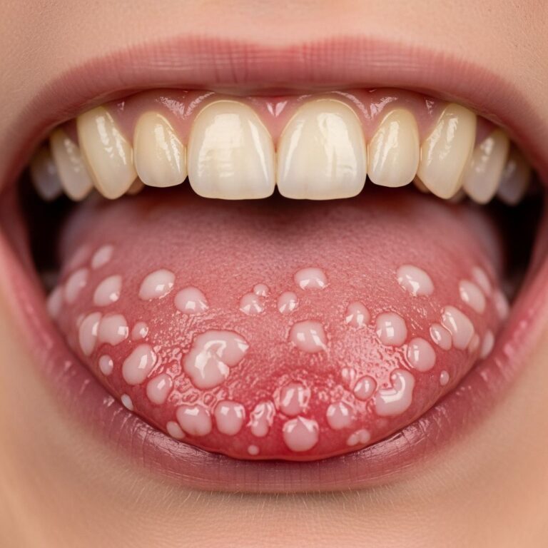
Mucosal Lichen Planus
Oral LP: Most common site—lacy white plaques on buccal mucosa, tongue, gingivae. Erosive form causes pain, scarring, 1-2% risk of squamous cell carcinoma.
Genital LP: Vaginal or vulval erosions in women; penile plaques in men. Symptomatic, requires biopsy to differentiate from lichen sclerosus.
Nail and Hair Involvement
Lichen planus of nails: Affects 10%; ridging, thinning, pterygium (scarring), onycholysis. Permanent scarring common.
Lichen planopilaris: Scarring alopecia with perifollicular erythema, scale. Variants: classic, frontal fibrosing alopecia (postmenopausal women), Graham-Little-Piccardi-Lassueur syndrome (scalp + body hair + non-scarring alopecia).
Lichenoid Drug Eruptions
Mimic idiopathic LP but often photosensitive or eczematous. Resolve 1-3 months after drug cessation; biopsy shows more eosinophils.
Other Lichenoid Dermatoses
| Condition | Key Features | Histology |
|---|---|---|
| Lichen nitidus | Minute white papules, trunk/limbs; self-limiting. | Granulomatous, claw-clipping of rete ridges. |
| Lichen aureus | Golden-brown patches on legs; persistent purpura. | Lichenoid infiltrate + hemosiderin. |
| Lichen striatus | Linear Blaschko lines, children; resolves 1-3 yrs. | Mixed spongiotic-lichenoid. |
| Annular lichenoid dermatitis of youth | Annular plaques on trunk; hypopigmented centers. | Lymphohistiocytic, vacuolar change. |
| Lichenoid actinic keratosis | Inflammatory papules on sun-damaged skin. | AK + lichenoid reaction. |
| Lichen planus pemphigoides | LP + bullae; autoantibodies to BP180. | Lichenoid + subepidermal blisters. |
| Lichenoid photodermatitis | Sun-exposed plaques; photoallergic. | Lichenoid pattern in photodamaged skin. |
Clinical Features
Symptoms: Pruritus (intense in hypertrophic), burning, pain (erosive/mucosal). Signs: Violaceous papules/plaques, Wickham striae (white lines, enhanced by iodine), nail dystrophy, scarring alopecia. Distribution: Flexors, oral/genital mucosa, nails.
Diagnosis
Clinical diagnosis in classic cases, confirmed by biopsy showing:
- Band-like lymphocytic infiltrate obscuring dermo-epidermal junction.
- Vacuolar basal degeneration, Civatte bodies.
- Hypergranulosis, saw-tooth rete ridges in LP.
Direct immunofluorescence: Fibrin + IgM at junction. Rule out mimics: Psoriasis, pityriasis rosea, secondary syphilis. Serology for HCV.
Treatment of Lichenoid Disorders
Treatment targets inflammation and symptoms; no cure.
Topical Therapies (First-line for localized)
- High-potency steroids (clobetasol) for skin; lower for face/mucosa.
- Calcineurin inhibitors (tacrolimus) for oral/genital.
- Intralesional steroids for hypertrophic/nail.
Systemic Therapies (Generalized/Severe)
- Oral glucocorticoids (prednisone 0.5-1mg/kg taper).
- Retinoids (acitretin), immunosuppressants (methotrexate, cyclosporine).
- Phototherapy (NB-UVB, PUVA) for cutaneous LP.
Specific Management
- Hep C-associated: Antivirals resolve LP.
- Drug-induced: Discontinue offender.
- Lichen planopilaris: Hydroxychloroquine, doxycycline.
- Mucosal: Monitor for malignancy.
Supportive: Emollients, antihistamines for itch. Refractory cases: Biologics (e.g., dupilumab experimental).
Frequently Asked Questions (FAQs)
Q: Is lichen planus contagious?
A: No, lichen planus and lichenoid disorders are not infectious; they are inflammatory/autoimmune.
Q: Can lichen planus cause cancer?
A: Erosive oral LP has 1-2% risk of squamous cell carcinoma; regular monitoring needed.
Q: How long does lichen planus last?
A: Cutaneous LP resolves in 1-2 years (80%); mucosal/nail can be chronic.
Q: What triggers lichen planus flares?
A: Stress, medications, trauma (Koebner), infections.
Q: Is lichen sclerosus a lichenoid disorder?
A: No, lichen sclerosus is distinct (thinning, sclerosis); though genital overlap, histologically different from LP.
References
- Lichens in dermatology — Indian Journal of Dermatology, Venereology and Leprology. 2017. https://ijdvl.com/lichens-in-dermatology/
- Lichenoid and interface dermatoses — PubMed/International Journal of Dermatology. 2017-05-01. https://pubmed.ncbi.nlm.nih.gov/28396069/
- Lichen Planus – Merck Manual Professional Edition — Merck Manuals. 2023. https://www.merckmanuals.com/professional/dermatologic-disorders/psoriasis-and-other-papulosquamous-disorders/lichen-planus
- Lichen Planus: What It Is, Causes, Types & Treatments — Cleveland Clinic. 2023-07-28. https://my.clevelandclinic.org/health/diseases/17723-lichen-planus
- Lichenoid disorders – DermNet — DermNet NZ. 2023. https://dermnetnz.org/topics/lichenoid-disorders
- Lichen planus and lichenoid dermatoses: Clinical overview — PubMed/Journal of the American Academy of Dermatology. 2018-10. https://pubmed.ncbi.nlm.nih.gov/30318136/
- Lichen planus – Symptoms and causes — Mayo Clinic. 2023-08-01. https://www.mayoclinic.org/diseases-conditions/lichen-planus/symptoms-causes/syc-20351378
Similar Articles


Read full bio of medha deb