Linear Focal Elastosis: Causes, Diagnosis, And Treatment Guide
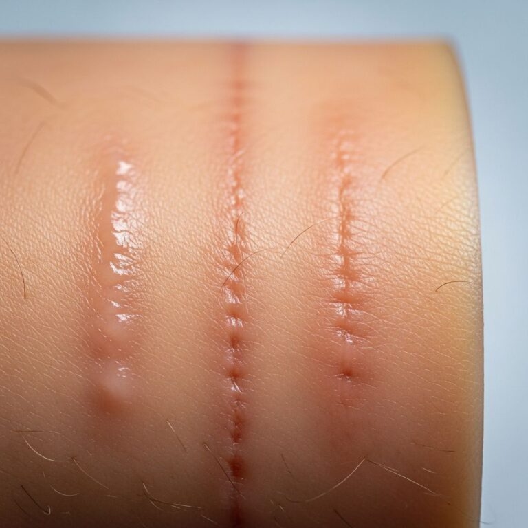
Uncommon dermal elastosis resembling stretch marks with yellow palpable lines on the lower back.

Linear focal elastosis is an uncommon form of dermal elastosis that resembles stretch marks.
Who gets linear focal elastosis?
Linear focal elastosis primarily affects young to middle-aged adults, with a notable predominance in males over females. It has been reported across various age groups, from children as young as 7 years to elderly individuals up to 89 years, without a specific racial predilection. The condition is rare, and its true prevalence remains unknown due to frequent misdiagnosis as more common stretch marks (striae distensae). Cases are often discovered incidentally during routine examinations, as the lesions are typically asymptomatic.
Although most commonly observed in the lumbosacral region of the back, linear focal elastosis can occur on the trunk, lower limbs, thighs, arms, breasts, and even the face in atypical presentations. Males are affected more frequently, possibly due to factors like rapid growth or physical activity, though no definitive demographic patterns are established beyond this male bias.
What causes linear focal elastosis?
The exact pathogenesis of linear focal elastosis remains unclear, but it is closely linked to alterations in dermal elastic tissue. A prominent hypothesis suggests it arises from a keloidal repair process (KRP) of elastic fibers damaged during the development of striae distensae (stretch marks). In several reported cases, linear focal elastosis lesions have followed or coexisted with striae distensae in the same anatomical distribution, showing transitional zones where atrophic striae evolve into hypertrophic, palpable bands.
Potential trigger factors include blunt trauma, pregnancy, significant weight loss, rapid growth during adolescence, or intense physical exercise. For instance, one case linked the condition to vigorous exercise, suggesting mechanical stress on the skin may promote excessive elastin regeneration. Unlike keloids, which involve transforming growth factor-beta (TGF-β) signaling, immunohistochemical studies in linear focal elastosis cases show negative TGF-β expression, indicating a distinct pathway for this abnormal elastogenesis.
Histopathologically, the condition features active elastogenesis, with elastic microfibrils connecting to fibroblast filaments, implying ongoing repair rather than de novo synthesis of malformed fibers. While not always associated with striae, the regenerative hypothesis is supported by electron microscopy revealing fragmented elastic tissue, microfibrillar components, and aggregated elastin.
Clinical features
Linear focal elastosis presents as asymptomatic, hypertrophic linear plaques that are yellow to red-brown in color, most commonly distributed symmetrically and horizontally across the lower and middle back in the lumbosacral area. These palpable bands are firm on touch, distinguishing them from the depressed, atrophic nature of striae distensae. Lesions are typically 1-3 mm wide and several centimeters long, running parallel to skin tension lines, and are often discovered incidentally without a history of trauma or symptoms like pain or itching.
- Distribution: Primarily lumbosacral back (90% of cases); less commonly trunk, abdomen, thighs, buttocks, arms, breasts, or face.
- Appearance: Yellowish palpable linear bands; may appear reddish in early stages.
- Symptoms: Asymptomatic; no pruritus, pain, or systemic associations.
- Progression: Stable once developed; may coexist with striae distensae, showing evolution from atrophic to indurated lesions.
Rare variants include diffuse or abdominal involvement, but the classic presentation aids clinical recognition. No associations with systemic diseases have been identified.
Differential diagnoses
The primary differential diagnosis is striae distensae (stretch marks), which are typically red (striae rubra) or white (striae alba), depressed/atrophic, and lack the hypertrophic, palpable quality of linear focal elastosis. Striae often follow pregnancy, obesity, or growth spurts, whereas linear focal elastosis bands are raised and yellow.
| Feature | Linear Focal Elastosis | Striae Distensae |
|---|---|---|
| Palpation | Palpable, hypertrophic | Depressed, atrophic |
| Color | Yellow to red-brown | Red or white |
| Elastic Tissue | Increased, elongated | Decreased |
| Location | Lumbosacral back | Variable (abdomen, thighs, etc.) |
Other considerations include:
- Pseudoxanthoma elasticum: Yellow papules/plaques with ruptured elastic fibers; systemic involvement (eyes, vessels).
- Connective tissue nevus: Firm nodules with excess collagen/elastin; often congenital.
- Elastofibroma: Thickened dermis with elastin aggregates; typically subscapular.
- Solar elastosis: Actinic damage on sun-exposed skin; wrinkled, not linear.
- Dermatofibrosis lenticularis disseminata: Hard yellow papules with perifollicular distribution.
Clinical distribution and biopsy distinguish linear focal elastosis.
Diagnosis
Diagnosis is primarily clinical, based on characteristic yellow palpable lines in the lumbosacral region. Skin biopsy confirms the condition, showing increased, elongated, wavy elastic fibers in the mid-to-deep dermis separating collagen bundles. Elastic stains (e.g., Verhoeff-Van Gieson) highlight the excess elastin, often clumped or fragmented, with possible early elastolysis. Electron microscopy reveals microfibrillar/granular components and active fibroblast involvement.
No routine lab tests or imaging are required, as there are no systemic features.
Treatment
No effective treatment exists for linear focal elastosis, as it is benign and asymptomatic. Topical corticosteroids and retinoids have shown no benefit. Cosmetic concerns may prompt observation or reassurance. Pulsed dye laser or other ablative therapies have been trialed anecdotally without consistent success. Management focuses on patient education regarding its harmless nature.
FAQs
Is linear focal elastosis the same as stretch marks?
No. Stretch marks (striae distensae) are atrophic and depressed, while linear focal elastosis features raised, yellow palpable bands due to excess elastic tissue.
Does linear focal elastosis require treatment?
No, as it is asymptomatic and benign. No proven therapies exist; observation is recommended.
Can linear focal elastosis appear anywhere on the body?
Primarily on the lower back, but reports exist on trunk, limbs, and face.
Is linear focal elastosis linked to other diseases?
No systemic associations; it is a localized dermal condition.
How is linear focal elastosis diagnosed?
Clinically by appearance; confirmed by biopsy showing excess dermal elastic fibers.
References
- Linear Focal Elastosis Following Striae Distensae — PubMed Central. 2011-12-01. https://pmc.ncbi.nlm.nih.gov/articles/PMC3229050/
- Linear focal elastosis — Cosmoderma. 2023-01-01. https://cosmoderma.org/linear-focal-elastosis/
- Linear focal elastosis — DermNet NZ. 2023-06-15. https://dermnetnz.org/topics/linear-focal-elastosis
- Linear focal elastosis — Clinical & Experimental Dermatology (Ovid). 2022-01-01. https://www.ovid.com/journals/cederm/fulltext/10.1093/ced/llac071~linear-focal-elastosis
- Linear focal elastosis associated with exercise — PubMed Central. 2017-01-01. https://pmc.ncbi.nlm.nih.gov/articles/PMC5288297/
- Linear Focal Elastosis: Histopathologic Diagnosis — Journal of Drugs in Dermatology. 2003-01-01. https://jddonline.com/articles/linear-focal-elastosis-histopathologic-diagnosis-of-an-uncommon-dermal-elastosis-S1545961603P0079X
Similar Articles


Read full bio of Sneha Tete