Livedo Reticularis: Causes, Symptoms, and Treatment
Understanding livedo reticularis: A comprehensive guide to this mottled skin condition.

Introduction to Livedo Reticularis
Livedo reticularis is a
mottled discoloration of the skin
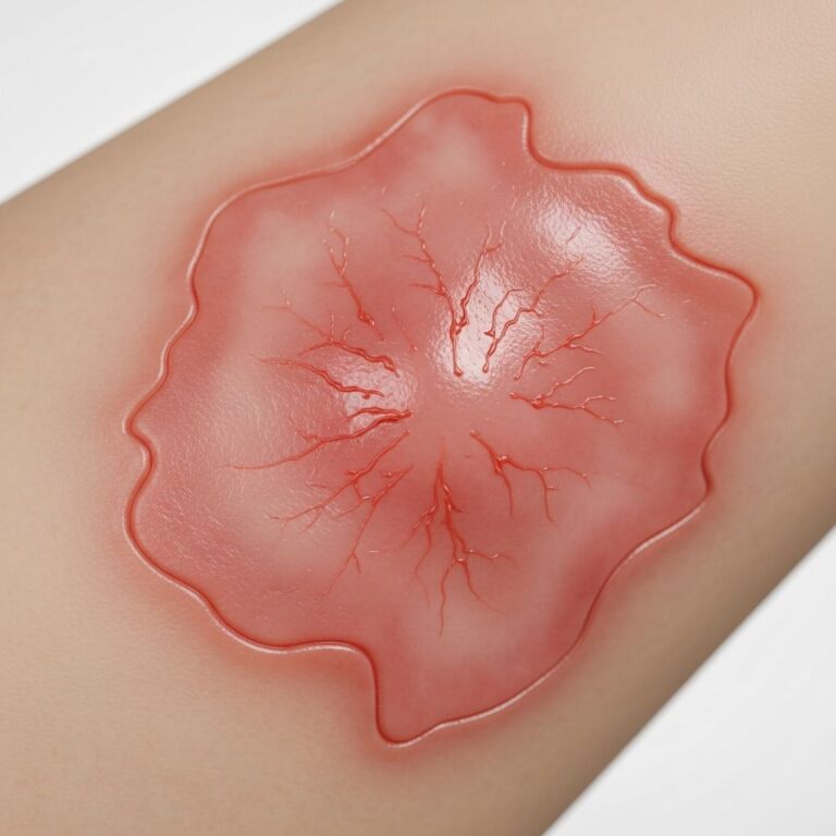
characterized by a distinctive net-like or lacy pattern that typically appears on the limbs, particularly the legs. The condition presents as a reddish-violet or bluish-purple reticulated pattern with clear borders, creating an appearance that resembles a fine lace network across the affected skin. This cutaneous manifestation results from disrupted blood flow in the small vessels beneath the skin surface, affecting how oxygen and nutrients are distributed to dermal tissues.Livedo reticularis is classified into two main types:
primary
andsecondary
. Primary livedo reticularis is typically benign and self-limited, often resolving without intervention. Secondary livedo reticularis, by contrast, indicates an underlying systemic condition requiring investigation and management. Understanding the distinction between these forms is crucial for determining appropriate treatment strategies and identifying potential health concerns.Pathophysiology and Mechanism
The development of livedo reticularis occurs through disruption of normal blood circulation in dermal arteries. This interruption can result from three primary mechanisms:
vascular spasm
,inflammation of blood vessel walls
, orvascular obstruction
. When blood flow is compromised in the small arteries and capillaries near the skin surface, the reduced oxygen delivery creates the characteristic discoloration pattern. The net-like appearance emerges because blood flow disruption occurs in a pattern that corresponds to the branching structure of dermal vasculature.In primary livedo reticularis, exposure to cold temperatures typically triggers the condition through reflex vasoconstriction of small blood vessels. This protective mechanism, normally designed to conserve body heat, can create the mottled appearance when the response is exaggerated or prolonged. Warming the affected area typically reverses the discoloration in primary cases.
Symptoms and Clinical Presentation
The
primary and most distinctive symptom
of livedo reticularis is the characteristic purple, net-like skin discoloration. This mottled pattern appears most commonly on the legs and lower extremities, though it can affect the face, trunk, buttocks, hands, and feet.Additional symptoms vary depending on the underlying cause and severity:
- Ongoing or persistent skin mottling that may worsen with cold exposure
- Cold or numb sensation in affected areas
- Skin discoloration that does not improve with warmth, suggesting secondary livedo reticularis
- Pain or development of skin ulcerations in severe cases linked with vascular diseases
- Reticulated erythema with fluctuating appearance that may come and go
In most cases of primary livedo reticularis,
no pain is present
, and the condition is purely cosmetic. However, if blood flow becomes completely blocked, pain and ulceration can develop, indicating the need for medical intervention. Secondary livedo reticularis may present with additional systemic symptoms related to the underlying condition, such as fever, joint pain, or other autoimmune manifestations depending on the causative disease.Causes of Livedo Reticularis
Primary Livedo Reticularis
Primary livedo reticularis typically results from benign environmental triggers, most commonly
cold exposure
. The condition is thought to be due to exaggerated spasms of blood vessels or disrupted blood flow regulation near the skin surface. This form is generally harmless and self-limited, often improving with age without requiring medical treatment.Secondary Livedo Reticularis
Secondary livedo reticularis is associated with various underlying systemic diseases and conditions affecting vascular integrity. These include:
- **Antiphospholipid syndrome** – an autoimmune condition affecting blood clotting
- **Systemic lupus erythematosus (lupus)** – an autoimmune inflammatory disease that can damage small blood vessels
- **Vasculitis** – inflammation of blood vessel walls
- **Cryoglobulinemia** – a condition involving cold-precipitating proteins in the blood
- **Infections** including hepatitis B, hepatitis C, and HIV
- **Malignancies** associated with hypercoagulable states
- **Acute pancreatitis** – inflammation of the pancreas causing secondary mottled skin as a rare symptom
- **Hypercoagulable states** and thrombotic disorders
Drug-Induced Livedo Reticularis
**Amantadine** is a well-known pharmaceutical cause of livedo reticularis, producing reddish-bluish lesions with a lacy, reticulated appearance typically on the trunk and legs. Other medications can similarly trigger this presentation. Importantly, amantadine-induced livedo reticularis usually resolves when the medication is discontinued. While typically asymptomatic aside from appearance, this drug-induced form can progress to ulceration if severe.
Diagnosis and Clinical Evaluation
**Clinical diagnosis** of livedo reticularis primarily relies on visual examination of the characteristic net-like skin pattern. However, establishing the underlying cause of secondary livedo reticularis requires comprehensive diagnostic workup.
Diagnostic Approach
Healthcare providers employ several diagnostic methods:
- **Physical examination** – assessment of the mottled pattern, extent, and associated skin findings such as nodules, retiform purpura, or necrosis
- **Detailed medical history** – including onset, duration, relationship to temperature changes, and associated systemic symptoms
- **Skin biopsy** – may be performed to evaluate histopathologic changes and rule out alternative diagnoses
- **Laboratory studies** – comprehensive testing to identify underlying systemic disease
Laboratory Investigation
To establish the cause of secondary livedo reticularis, the following laboratory studies may be indicated:
- Anticardiolipin antibody testing
- Antiphospholipid antibody testing
- Autoimmune panel
- Coagulation panel
- Complete blood count
- Cryoprotein testing
- Hepatitis B and C serology
- HIV serology
- Kidney and liver function tests
- Lupus anticoagulant panel
- Serum protein electrophoresis
Assessment of Course and Severity
Determining whether livedo reticularis is acute, chronic, or fulminant guides treatment decisions and urgency of evaluation. Identification of other cutaneous signs including nodules, retiform purpura, or necrosis suggests more serious underlying pathology requiring prompt intervention.
Differential Diagnosis
Several other dermatologic conditions present similarly to livedo reticularis and must be distinguished during evaluation:
- **Erythema ab igne** – thermal injury pattern from heat exposure
- **Erythema infectiosum** – fifth disease with characteristic erythema
- **Poikiloderma** – combination of atrophy, dyspigmentation, and telangiectasia
- **Mycosis fungoides** – cutaneous T-cell lymphoma
- **Dermatomyositis** – autoimmune inflammatory myopathy with skin manifestations
- **Graft versus host disease** – immune reaction following bone marrow transplantation
- **Reticulated erythematosus mucinosis** – rare dermatologic condition
Treatment Options
Primary Livedo Reticularis Treatment
**Medical treatment is generally not indicated** for primary livedo reticularis, as symptoms often resolve with age and the condition remains benign. However, for patients bothered by the cosmetic appearance, several therapeutic options exist.
Nonpharmacologic Management
Conservative measures form the foundation of primary livedo reticularis management:
- **Keeping warm** – maintaining adequate body temperature, particularly protecting the legs from cold exposure
- **Avoiding smoking** – which can impair circulation and worsen symptoms
- **Stress reduction** – avoiding stressful situations that may trigger or exacerbate symptoms
- **Cold avoidance** – preventing exposure to chilling temperatures that trigger vasoconstriction
Pharmacologic Options
When nonpharmacologic measures are insufficient, medications targeting vascular function may be considered:
- **Nifedipine** – a calcium channel blocker that promotes vasodilation
- **Pentoxifylline** – improves blood flow and rheologic properties
Secondary Livedo Reticularis Treatment
**Treatment of secondary livedo reticularis depends fundamentally on addressing the underlying disease**. Improvement in skin appearance typically follows successful management of the causative condition.
Disease-Specific Approaches
For conditions involving hypercoagulability or thrombotic disease:
- **Anticoagulation therapy** – blood thinning medications may be recommended when blood clots are the primary problem
- **Low-dose aspirin therapy** – for antiplatelet effects
- **Warfarin** – for more intensive anticoagulation when indicated
Alternative Therapies for Resistant Cases
When standard approaches prove insufficient, additional therapeutic modalities may be effective:
- **Hyperbaric oxygen therapy** – enhances tissue oxygenation to improve perfusion
- **Psoralen with ultraviolet A (PUVA) therapy** – photochemotherapy may improve appearance in resistant cases
- **Pentoxifylline** – particularly useful for improving microcirculation
When to Seek Medical Care
While livedo reticularis often requires no treatment, certain circumstances warrant evaluation by a healthcare provider:
- The discolored, mottled skin does not resolve with warming, suggesting secondary disease
- Mottled skin appears alongside other concerning symptoms
- Painful lumps develop in affected skin
- Ulcerations or sores develop in the discolored areas
- You have a condition affecting blood flow in your limbs
- New skin symptoms develop and you have a connective tissue disease
Prognosis and Prevention
The prognosis for livedo reticularis varies significantly by type. Primary livedo reticularis typically has an excellent prognosis, with the condition often resolving spontaneously or remaining stable as a benign cosmetic concern. Secondary livedo reticularis prognosis depends on successful management of the underlying systemic condition.
**Prevention strategies** differ by type. Primary livedo reticularis can often be prevented or minimized by avoiding cold exposure and maintaining adequate warmth. Secondary livedo reticularis prevention may be challenging but requires close adherence to care instructions for the underlying condition affecting blood flow and vascular integrity.
Frequently Asked Questions
Q: Is livedo reticularis dangerous?
A: Primary livedo reticularis is harmless and purely cosmetic. Secondary livedo reticularis, however, may indicate a serious underlying condition requiring medical evaluation and treatment of the causative disease.
Q: Will livedo reticularis go away on its own?
A: Primary livedo reticularis often resolves without treatment, particularly with age. Secondary livedo reticularis typically improves when the underlying condition is successfully managed.
Q: Does warming the skin help livedo reticularis?
A: Keeping warm, especially the legs, may relieve skin discoloration in primary livedo reticularis. However, if discoloration persists despite warming, this suggests secondary livedo reticularis and warrants medical evaluation.
Q: What medications cause livedo reticularis?
A: Amantadine is the most well-known pharmaceutical cause, producing lacy red-blue lesions that resolve upon medication discontinuation. Other medications may occasionally cause this presentation.
Q: How is livedo reticularis diagnosed?
A: Diagnosis is primarily clinical based on the characteristic appearance. Blood tests and skin biopsy may be performed to evaluate for underlying systemic disease in secondary cases.
References
- Mottled Skin (Livedo Reticularis): Causes, Symptoms, and Diagnosis — Cleveland Clinic. 2024. https://my.clevelandclinic.org/health/symptoms/24429-mottled-skin
- Diagnosis and Treatment of Livedo Reticularis on the Legs — Actas Dermo-Sifiliográficas. 2008. https://www.actasdermo.org/en-diagnosis-treatment-livedo-reticularis-on-articulo-S1578219008703275
- Livedo Reticularis – Dermatology Advisor — Dermatology Advisor. 2024. https://www.dermatologyadvisor.com/ddi/livedo-reticularis/
- Livedo Reticularis: When is it a concern? — Mayo Clinic. 2024. https://www.mayoclinic.org/diseases-conditions/vasculitis/expert-answers/livedo-reticularis/faq-20057864
- Livedo Reticularis — MedlinePlus Medical Encyclopedia. 2024. https://medlineplus.gov/ency/article/001478.htm
- Mottled Skin (Livedo Reticularis): Causes, Treatment, and More — Medical News Today. 2024. https://www.medicalnewstoday.com/articles/321422
Similar Articles


Read full bio of medha deb