Livedoid Vasculopathy: Clinical Features, Diagnosis & Management
Complete guide to livedoid vasculopathy: understanding symptoms, diagnostic approaches, and therapeutic strategies.

Livedoid Vasculopathy: Understanding a Rare Vascular Disorder
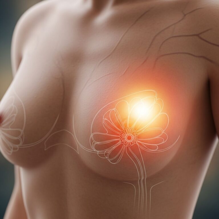
Livedoid vasculopathy (LV) is a rare, chronic thrombotic dermal condition characterized by recurrent, painful ulcers primarily affecting the lower limbs. This uncommon vascular disorder involves the occlusion of dermal blood vessels, resulting in a distinctive clinical presentation that combines livedo patterns, painful ulceration, and characteristic scarring. Understanding the clinical features, diagnostic criteria, and management strategies is essential for healthcare providers and patients affected by this debilitating condition.
Clinical Features and Presentation
Livedoid vasculopathy presents with a characteristic clinical triad consisting of three distinctive features:
- Livedo racemosa or livedo reticularis — a lacy, netlike pattern of red to purplish skin discoloration that creates a retiform appearance on affected areas
- Painful ulcers — excruciating, recurrent lesions that develop over months to years, typically localized to the lower extremities
- Atrophie blanche — porcelain-white, star-shaped scars that remain after ulcers heal, representing permanent cutaneous changes
The initial manifestations of livedoid vasculopathy typically involve clusters of painful, flat or raised spots arranged in star-shaped patterns around the ankle region. These lesions appear red or purplish in color and primarily affect the skin surrounding both ankles, though they may extend to the upper portions of the feet and shins. The condition predominantly involves bilateral lower limb involvement, though unilateral presentations have been documented in rare cases.
Livedo racemosa usually appears before ulceration occurs and represents the initial clinical manifestation of dermal vascular occlusion. The painful ulcers that subsequently develop can persist for extended periods and are accompanied by significant morbidity due to their intensity. Studies demonstrate that livedoid vasculopathy causes intensive pain, with median pain scores reaching 65.0 on the visual analogue scale, leading to dramatic impairment of quality of life and functional capacity.
Atrophie blanche refers to the characteristic whitish scars that appear as ulcers heal. These scars are also star-shaped and represent permanent markers of previous ulceration. The development of these scars indicates successful wound healing but serves as visual evidence of the disease’s chronicity and severity.
Atypical and Associated Symptoms
Less common manifestations of livedoid vasculopathy include bluish discoloration of the skin, numbness in the feet, and skin hardening of the lower legs. Some patients experience symptoms suggestive of polyneuropathy, including abnormal sensations (dysesthesia and hypoesthesia) predominantly located on the outer aspects of the lower legs and the back of the foot. These neurological symptoms may occasionally be associated with mononeuritis multiplex, a form of peripheral neuropathy.
Underlying Pathophysiology
Livedoid vasculopathy is currently classified as a coagulation disorder rather than an inflammatory vasculitis, distinctly differentiated from conventional vasculitic conditions. The disease process involves recurrent focal, non-inflammatory thrombosis of the superior superficial and medium dermal venules, particularly on the lower extremities and bilaterally. The vaso-occlusion of the cutaneous microcirculation manifests histopathologically as hyalinization of blood vessel walls leading to fibrin thrombi and vessel occlusion.
Although evidence suggests hypercoagulability and secondary inflammatory changes contribute to the pathology, the complete molecular mechanisms underlying livedoid vasculopathy remain incompletely understood. The condition results in skin ischemia due to occlusion of dermal vessels, which directly triggers the intense pain experienced by affected patients. Blood and pressure accumulation in the dermal superficial veins contributes to the livedo patterns observed clinically.
Diagnosis and Diagnostic Investigations
Livedoid vasculopathy is fundamentally a clinical diagnosis supported and confirmed by skin biopsy findings. The diagnosis should be made based on a synthesis of clinical presentation, patient history, physical examination findings, and histopathological confirmation for appropriate exclusion of differential diagnoses.
Clinical and Historical Assessment
A thorough clinical history, detailed dermatological examination, and comprehensive review of systems form the foundation of diagnostic evaluation. Healthcare providers should inquire about the chronicity of symptoms, progression pattern, associated pain characteristics, and any systemic manifestations. The clinical appearance of livedoid vasculopathy is often quite typical, featuring the characteristic triad of livedo patterns, painful ulcers, and atrophie blanche scars.
Notably, livedoid vasculopathy follows a chronic time course with lesions often developing for months before a definitive diagnosis is established. In clinical case series, the median interval between symptom onset and diagnosis was approximately 10 months, with a median interval of 22.5 months between symptom onset and initiation of treatment. This diagnostic delay underscores the importance of clinical awareness and appropriate use of confirmatory investigations.
Skin Biopsy and Histopathology
A skin biopsy is necessary to confirm the diagnosis of livedoid vasculopathy and should be obtained from a red papule or the edge of a newly developed ulcer. The most suitable biopsy technique is a fusiform incisional biopsy that includes subcutaneous fat, allowing for assessment of lesions at multiple dermal levels.
Characteristic histopathological findings of livedoid vasculopathy include:
- Hyalinization and thickened blood vessel walls with a glassy appearance to some cells (hyaline degeneration)
- Fibrin deposition within vessel walls, which can be difficult to identify on routine staining
- Intraluminal hyaline thrombi in blood vessels, particularly in the upper and middle dermis (high frequency during acute phase)
- Endothelial proliferation and increased endothelial cells forming the inner lining of blood vessels
- Blood clots (thrombi) within vessels
- Vascular occlusion by thrombosis
- Minimal inflammation with sparse perivascular inflammatory infiltrate
- Leukocytoclasia may be observed in the acute phase but is not essential for diagnosis
Immunofluorescence and Laboratory Investigations
Direct immunofluorescence studies often reveal deposition of immunoglobulin and complement components throughout the superficial, mid-dermal, and deep dermal vasculature, though these findings are non-diagnostic and serve primarily as supporting evidence.
General investigations should include comprehensive laboratory assessment, though in many cases of livedoid vasculopathy, test results remain normal. Thrombophilia laboratory testing is recommended for all patients to identify potential underlying coagulation disorders. Additional testing may be necessary to determine whether any thrombophilia is inherited or acquired, including evaluation of coagulating factors and their specific mutations.
Transcutaneous oximetry demonstrates reduced oxygen flow in most patients with livedoid vasculopathy, reflecting the vascular insufficiency underlying the condition. Imaging studies for peripheral vascular disease may also be performed to evaluate for concurrent microvascular compromise.
Differential Diagnosis
Because the signs and symptoms of livedoid vasculopathy overlap with those of other diseases, careful differential diagnosis is essential. The primary conditions to be excluded include:
- Chronic venous insufficiency — caused by dysfunctional vein valves that prevent proper blood return to the heart, resulting in leg swelling and similar skin symptoms
- Peripheral arterial disease — characterized by reduced arterial blood flow to the extremities
- Vasculitis — inflammatory vascular disorders that can mimic livedoid vasculopathy but represent distinct pathophysiologic processes
- Cutaneous polyarteritis nodosa (cutaneous PAN) — a small- to medium-vessel necrotizing vasculitis affecting the deep dermis and hypodermis with comparable clinical symptoms
- Sneddon’s syndrome — characterized by livedo racemosa with systemic involvement, particularly neurological manifestations
Ankle-brachial index testing, which compares blood pressure in the ankle with blood pressure in the arm, assists in diagnosing peripheral artery disease. Doppler ultrasound studies help distinguish peripheral artery disease and chronic venous insufficiency from livedoid vasculopathy by demonstrating specific vascular flow abnormalities characteristic of these alternative diagnoses.
The presence of livedo racemosa frequently triggers patient referrals to dermatology for evaluation and exclusion of Sneddon’s syndrome. Currently, there is no evidence that livedoid vasculopathy affects organs beyond the skin. Patients presenting with neurological findings alongside livedoid vasculopathy warrant referral to neurologists for comprehensive evaluation of concurrent neurological conditions.
Current Diagnostic Approach
At present, no validated diagnostic score exists for livedoid vasculopathy. Diagnosis relies upon clinical judgment synthesizing historical information, examination findings, and histopathological confirmation. This synopsis of clinical and histological findings enables healthcare providers to establish definitive diagnosis while excluding alternative diagnoses with overlapping presentations.
Therapeutic Management and Treatment Goals
The primary therapeutic objective in managing livedoid vasculopathy is to reduce pain intensity, minimize ulceration, and prevent the development or progression of atrophie blanche. Early and adequate therapy is essential for maintaining quality of life and avoiding irreversible complications associated with chronic ulceration and scarring.
Current management approaches include anticoagulation therapy, antiplatelet agents, vasodilators, and various adjunctive treatments aimed at improving microvascular circulation and reducing thrombotic episodes. The heterogeneous nature of livedoid vasculopathy and incomplete understanding of its molecular mechanisms necessitate individualized treatment approaches tailored to patient-specific factors and disease severity.
Clinical Course and Chronicity
Livedoid vasculopathy typically follows a chronic, recurrent course extending over years to decades. The delayed interval between initial symptom manifestation and diagnosis represents a significant clinical challenge, as patients often endure prolonged pain and progressive tissue damage before receiving appropriate treatment. The condition is rare in children, with most cases presenting in adults.
The chronic nature of livedoid vasculopathy, combined with the intensity of associated pain and significant quality-of-life impairment, underscores the importance of early recognition, prompt diagnosis, and initiation of appropriate therapeutic interventions.
Frequently Asked Questions
Q: Is livedoid vasculopathy curable?
A: Livedoid vasculopathy is a chronic condition without a known cure. Management focuses on controlling symptoms, reducing pain, minimizing ulceration, and preventing further atrophie blanche development. Early intervention can help maintain quality of life and prevent irreversible complications.
Q: What causes livedoid vasculopathy?
A: The exact cause is not completely understood, but livedoid vasculopathy is classified as a coagulation disorder involving thrombosis of dermal blood vessels. It appears to involve hypercoagulability and secondary inflammation, though the complete molecular mechanisms require further investigation.
Q: How long does diagnosis typically take?
A: The median interval between symptom onset and diagnosis is approximately 10 months, with an average of 22.5 months before treatment begins. This diagnostic delay emphasizes the importance of clinical awareness among healthcare providers.
Q: Can livedoid vasculopathy affect organs other than skin?
A: Currently, there is no evidence that livedoid vasculopathy affects organs beyond the skin. However, some patients may experience associated peripheral neuropathy symptoms, warranting evaluation by specialists.
Q: How is livedoid vasculopathy different from vasculitis?
A: Livedoid vasculopathy is classified as a coagulation disorder rather than an inflammatory vasculitis. It involves thrombotic occlusion of dermal vessels with minimal inflammation, distinctly differentiating it from conventional vasculitic conditions.
References
- Livedoid Vasculopathy — Symptoms, Causes, Treatment — National Organization for Rare Disorders (NORD). https://rarediseases.org/rare-diseases/livedoid-vasculopathy/
- Livedoid Vasculopathy — DermNet. https://dermnetnz.org/topics/livedoid-vasculopathy
- Livedoid Vasculopathy – A Diagnostic and Therapeutic Challenge — Frontiers in Medicine, 2022. https://www.frontiersin.org/journals/medicine/articles/10.3389/fmed.2022.1012178/full
- Livedoid Vasculopathy — StatPearls, National Center for Biotechnology Information (NCBI). https://www.ncbi.nlm.nih.gov/books/NBK559037/
- Livedoid Vasculopathy — VisualDx Medical Reference — VisualDx. https://www.visualdx.com/visualdx/diagnosis/livedoid+vasculopathy
Similar Articles


Read full bio of Sneha Tete