Lymphocytoma Cutis: Essential Guide To Diagnosis And Treatment
Comprehensive guide to lymphocytoma cutis: causes, diagnosis, treatment, and differentiation from lymphoma.

Lymphocytoma cutis, also known as cutaneous lymphoid hyperplasia or pseudolymphoma, is a rare benign skin condition characterized by an exaggerated immune response leading to localized proliferation of lymphocytes in the skin. It presents as solitary or multiple firm nodules, typically on the face, ears, or limbs, mimicking malignant lymphoma but remaining reactive and polyclonal. This condition arises from various triggers including infections, arthropod bites, medications, and vaccinations, requiring careful clinicopathological correlation for diagnosis.
What is lymphocytoma cutis?
Lymphocytoma cutis represents a pseudolymphomatous reaction pattern in the skin, where lymphoid follicles and infiltrates accumulate without malignant transformation. It is not a true neoplasm but a hyperplastic response to antigenic stimuli. Clinically, it manifests as soft, doughy, or firm nodules ranging from skin-coloured to red-brown or red-purple hues, often 1–3 cm in size. These lesions can be solitary or grouped, predominantly affecting the head, neck, upper extremities, and earlobes.
The term ‘lymphocytoma cutis’ encompasses both B-cell and T-cell predominant forms, with B-cell types being more common and featuring germinal centres. Unlike lymphoma, it lacks monoclonality and shows polyclonality on gene rearrangement studies. Spontaneous resolution may occur upon removal of the inciting agent, but persistent cases necessitate intervention.
Who gets lymphocytoma cutis?
Lymphocytoma cutis affects individuals across all ages but shows a predilection for children and young adults, particularly in cases linked to infections like Borrelia. Females may be slightly more prone, especially for earlobe lesions associated with piercings. Immunocompetent hosts are primarily impacted, though immunosuppression can exacerbate presentations. Geographic variations exist, with Borrelia-associated cases more common in Europe.
- Children: Often post-vaccination or insect bites.
- Adults: Drug-induced or idiopathic.
- Earlobe predilection in piercing-related cases.
What causes lymphocytoma cutis?
The aetiology is multifactorial, stemming from hypersensitivity reactions to diverse stimuli:
- Infections: Borrelia burgdorferi (Lyme disease), herpes zoster, varicella, HIV, hepatitis C, Leishmania.
- Arthropod assaults: Mosquito, tick, spider bites, scabies.
- Drugs: Antidepressants (mirtazapine, fluoxetine), antihypertensives (amlodipine), anticonvulsants, gold therapy.
- Vaccinations: COVID-19, HPV, hepatitis B.
- Foreign bodies: Piercings, tattoos, acupuncture.
- Physical factors: Trauma, UV light, cold exposure.
- Idiopathic cases comprise up to 30%.
Borrelial lymphocytoma cutis, a specific subtype, presents as bluish-red nodules on the earlobe or areola, responding to antibiotics targeting the infection.
What are the clinical features of lymphocytoma cutis?
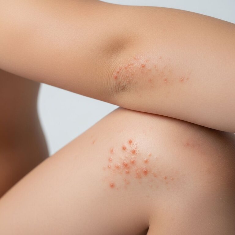
Lesions appear as painless, slowly enlarging nodules or plaques:
- Solitary: Common on face (cheek, eyelid), ears, nipples, scrotum.
- Multiple: Grouped on limbs or trunk.
- Size: 0.5–3 cm, firm or doughy consistency.
- Colour: Erythematous, violaceous, red-brown; may ulcerate rarely.
- Symptoms: Asymptomatic or mildly pruritic; no systemic signs.
Figure 1: Red nodule on cheek; Figure 2: Multiple earlobe nodules (descriptive based on typical presentations).
Diagnosis
Skin biopsy is essential to confirm the benign nature and exclude lymphoma. Histology reveals:
- Dense nodular lymphocytic infiltrate in dermis/subcutis.
- Well-formed germinal centres with tingible body macrophages.
- Polyclonal B-cells (kappa/lambda ratio normal), mantle zones, eosinophils, plasma cells.
- B:T cell ratio <3:1; CD20+, CD79a+ B-cells; Bcl-2 negative in germinal centres.
Immunohistochemistry and PCR for immunoglobulin/TCR gene rearrangements confirm polyclonality. Blood tests screen for infections (Borrelia serology, HIV), and imaging rules out systemic disease.
Differential diagnosis
Lymphocytoma cutis mimics cutaneous B-cell lymphomas:
| Feature | Lymphocytoma Cutis | Cutaneous Lymphoma |
|---|---|---|
| Germinal centres | Well-formed, tingible bodies present | Ill-formed, expanded |
| Proliferation rate | High in follicles | Low |
| Bcl-2 | Negative in centres | Positive |
| Light chains | Polyclonal | Monoclonal |
| CD10/Bcl6 | Follicle-restricted | Interfollicular |
Other differentials: Basal cell carcinoma, adnexal tumours, sarcoidosis, lupus.
Treatment of lymphocytoma cutis
No standardized guidelines exist; management targets aetiology and lesion extent:
Conservative
- Remove trigger (stop drug, treat infection).
- Observation: 20–50% spontaneous regression.
Localized disease
- Intralesional steroids (triamcinolone 10–20 mg/mL): 70–90% response.
- Excision or curettage.
- Laser (pulsed dye, CO2), cryotherapy.
- Topical tacrolimus, imiquimod.
Widespread/generalized
- Oral hydroxychloroquine (6.5 mg/kg/day).
- Thalidomide, interferon-alpha.
- Mycophenolate mofetil for recalcitrant cases.
- Phototherapy (PUVA, UVA1).
Borrelial cases: Doxycycline or penicillin first-line. Recurrence post-treatment is common; monitor clinically.
What is the outcome for lymphocytoma cutis?
Benign with excellent prognosis; no malignant potential. Resolution occurs in most cases with appropriate therapy, though persistence or relapse may require escalation. Long-term follow-up advised to detect rare progression mimics.
Related topics
- Cutaneous B-cell lymphoma
- Borrelial lymphocytoma
- Drug-induced pseudolymphoma
- Jessner-Kanof lymphocytic infiltrate
Frequently asked questions
Is lymphocytoma cutis cancerous?
No, it is a benign reactive hyperplasia, distinguished from lymphoma by polyclonality and reactive features.
Does it go away on its own?
Yes, often after trigger removal, but treatment accelerates resolution.
How is it treated?
Intralesional steroids for localized; antimalarials or immunosuppressants for widespread.
Can it recur?
Yes, especially if trigger persists; re-biopsy if changes occur.
Is biopsy always needed?
Yes, to rule out lymphoma.
References
- Recalcitrant lymphocytoma cutis successfully treated with mycophenolate mofetil — J Cutan Med Surg. 2021-06-15. https://pmc.ncbi.nlm.nih.gov/articles/PMC8207277/
- Lymphocytoma Cutis – A Case Report — Skin. 2023. https://skin.dermsquared.com/skin/article/view/2321
- Lymphocytoma cutis — Plastic Surgery Key. 2024. https://plasticsurgerykey.com/lymphocytoma-cutis/
- Lymphocytoma cutis (cutaneous lymphoid hyperplasia) — DermNet NZ. 2024-01-01. https://dermnetnz.org/topics/lymphocytoma-cutis
- Treatment of Cutaneous Pseudolymphoma: A Systematic Review — Acta Derm Venereol. 2020-10-22. https://www.medicaljournals.se/acta/content/html/10.2340/00015555-2841
Similar Articles


Read full bio of medha deb