Lymphomatoid Papulosis Pathology: 6 Key Histological Subtypes
Comprehensive pathology of lymphomatoid papulosis: histological features, subtypes, and diagnostic insights for clinicians.

Lymphomatoid papulosis (LyP) represents a distinctive clinicopathological entity within the spectrum of primary cutaneous CD30-positive lymphoproliferative disorders. Characterised by recurrent, self-resolving papulonodular eruptions, LyP exhibits malignant histological features despite its indolent clinical behaviour. This article delineates the pathological hallmarks, subtype classifications, immunohistochemical profiles, molecular characteristics, and diagnostic considerations essential for pathologists and dermatologists.
Histological Features
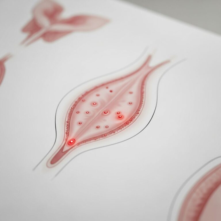
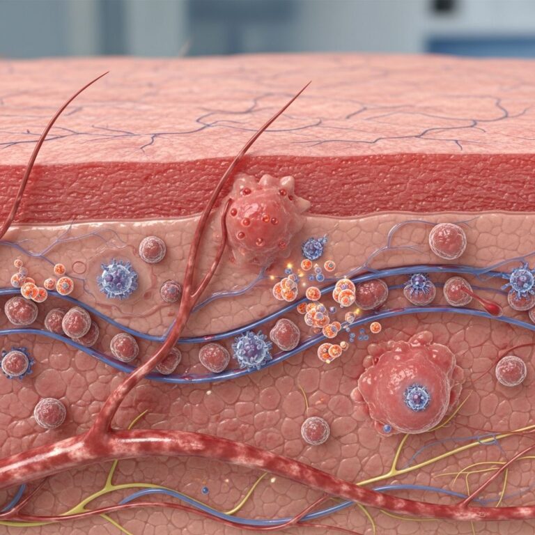
The histopathology of LyP is defined by a polymorphous infiltrate dominated by atypical CD30-positive large lymphoid cells within a background of inflammatory cells. Early lesions (<48 hours) show wedge-shaped epidermal hyperplasia with superficial perivascular lymphocytic infiltration, evolving into prominent papillary dermal oedema by days 3-5. Mature lesions (5-10 days) feature dense, wedge-shaped dermal infiltrates extending into the deep dermis or subcutis, comprising small lymphocytes, histiocytes, eosinophils, and scattered neutrophils alongside hallmark large atypical cells with pleomorphic, hyperchromatic nuclei, irregular nuclear membranes, and abundant cytoplasm.
These atypical cells, often binucleate or multinucleate, constitute 5-100% of the infiltrate and cluster around vessels or adnexa. Epidermotropism is common, with atypical cells migrating singly or in small groups into the epidermis, sometimes forming Pautrier-like microabscesses. Ulceronecrotic lesions display extensive epidermal necrosis with overlying fibrinopurulent exudate and dermal haemorrhage. Spontaneous regression is marked by interface dermatitis, basal vacuolisation, and apoptosis, culminating in fibrosis and scarring.
Subtypes of Lymphomatoid Papulosis
LyP is classified into six histological subtypes (A-F), each with unique morphological features, though clinical presentation remains consistent across variants.
Type A (Classical, 80% of cases)
The most prevalent subtype mirrors systemic anaplastic large cell lymphoma (ALCL). It features scattered to cohesive sheets of large CD30+ cells (>15 microns, 25-100% of infiltrate) with hallmark cells, multinucleate Reed-Sternberg-like forms, and inflammatory background including eosinophils and histiocytes. Mitoses are frequent.
Type B
Resembling primary cutaneous CD30- mycosis fungoides, type B shows epidermotropic small to medium-sized atypical lymphocytes with cerebriform nuclei (type B1) or larger cells with less epidermotropism (type B2). CD30 expression is weak or absent.
Type C
Characterised by monotonous sheets of large anaplastic CD30+ cells (>75% of infiltrate) with minimal inflammatory background, closely mimicking ALCL but distinguished by spontaneous regression.
Type D
Features epidermotropic small to medium CD8+ atypical lymphocytes with granular cytoplasm, resembling primary cutaneous aggressive epidermotropic CD8+ cytotoxic T-cell lymphoma. CD30 positivity is variable.
Type E
Angioinvasive pattern with individually necrotic CD8+ CD30+ large atypical cells dissecting collagen and occluding vessels, associated with rapidly progressive ulceration.
Type F
Recently described, shows epidermotropic CD30+ large atypical cells without angioinvasion, distinguished from type D by CD30 expression and lack of granzyme B.
Type P (Poikilodermatous)
Rare variant with interface dermatitis, epidermal atrophy, and telangiectasias, featuring CD30+ atypical cells in a lichenoid infiltrate.
Immunohistochemistry
Immunophenotyping is crucial for diagnosis. Classical type A LyP cells express CD30 diffusely (membranous/cytoplasmic), CD3, CD4, CD45RO, and cytotoxic proteins (granzyme B, perforin, TIA-1). CD8 is negative. CD20/B-cell markers are absent in atypical cells; CD15, CD30 variable in neutrophils/histiocytes.
Type B typically shows CD3+, CD4+, CD30-/weak+. Type D/E/F express CD8+, CD30+, cytotoxic markers. CD56 occasionally positive in type D. Loss of pan-T-cell markers (CD2, CD5, CD7) supports malignancy.
| Subtype | Key IHC Markers | Background |
|---|---|---|
| A | CD30+, CD4+, cytotoxic+ | Mixed inflammatory |
| B | CD4+, CD30-/weak | Epidermotropic small cells |
| C | CD30+ sheets | Paucicellular |
| D | CD8+, CD30+, cytotoxic+ | Granular cytoplasm |
| E | CD8+, CD30+, angioinvasive | Necrotic cells |
Molecular Findings
Clonal T-cell receptor (TCR) gene rearrangements are detected in 60-90% of cases via PCR, supporting neoplastic nature despite benign behaviour. Identical clones may link LyP to associated lymphomas. Next-generation sequencing reveals recurrent mutations: NF-κB pathway (TNFAIP3 44%, IKBKB 18%), JAK/STAT (STAT3/5B 40%), epigenetic regulators (TET2, CREBBP), and TP53. FAT1, FAT4 mutations noted. Type D frequently harbours TP63 rearrangements. DUSP22, MYC rearrangements rare.
Electron Microscopy
Ultrastructurally, atypical cells display irregular, convoluted nuclei with marginated chromatin, prominent nucleoli, and abundant cytoplasm with Golgi apparatus, rough endoplasmic reticulum, and lysosomes. No Birbeck granules.
Pathogenesis
Though aetiology unknown, LyP involves clonal expansion of CD30+ activated T-cells with defective apoptosis. CD30 signalling promotes survival; chronic antigenic stimulation or viral triggers (HTLV-1, EBV rare) implicated. Peripheral blood T-cell clonality observed in some.
Clinical Correlation
Lesions present as crops of erythematous papules (2-20mm) on trunk/extremities, evolving to haemorrhagic/necrotic crusts over 2-12 weeks with varioliform scarring. Average age 45 years; male predominance (M:F 1.5:1). Incidence 1.2-1.9/million.
Complications and Associations
Despite indolent course (100% 5-year survival), 15-25% develop secondary lymphomas: mycosis fungoides (most common), pcALCL, Hodgkin lymphoma, systemic ALCL. Risk factors: older age, monoclonal TCR. Quality-of-life impacted by scarring, ulceration.
Differential Diagnosis
- Primary cutaneous ALCL: Larger persistent plaques/tumours, contiguous infiltrates, higher CD30 expression.
- Mycosis fungoides: Patch/plaque stage, epidermotropism without large CD30+ cells.
- Pityriasis lichenoides: Interface changes, fewer atypical cells.
- Arthropod bites: Eosinophil-rich, no clonality.
Diagnostic Approach
Requires clinicopathological correlation: multiple biopsies (early/mature), large (4-6mm punch). Exclude systemic disease (CT/PET, marrow if B-symptoms, abnormal labs). TCR clonality aids.
Treatment Considerations
No cure; manage flares with skin-directed therapies (topical steroids, phototherapy), low-dose methotrexate (effective), PUVA. Monitor for lymphoproliferative progression.
Frequently Asked Questions
What is the hallmark histological finding in lymphomatoid papulosis?
Atypical CD30-positive large lymphoid cells in a polymorphous inflammatory infiltrate with epidermotropism.
How reliable is TCR clonality for diagnosis?
Present in 60-90% but not diagnostic alone; must correlate clinically.
Which LyP subtype mimics ALCL most closely?
Type C, with sheets of monotonous CD30+ cells.
Is systemic staging required at diagnosis?
Not routinely unless extracutaneous symptoms present.
What is the risk of progression to lymphoma?
15-25%, primarily mycosis fungoides or pcALCL.
References
- Lymphomatoid Papulosis Patient Information — Newcastle Hospitals NHS Foundation Trust. 2023. https://www.newcastle-hospitals.nhs.uk/services/dermatology/patient-dermatology-information-leaflets/lymphomatoid-papulosis/
- Lymphomatoid Papulosis — DermNet NZ. 2024-01-15. https://dermnetnz.org/topics/lymphomatoid-papulosis
- Lymphomatoid Papulosis — StatPearls [Internet]. NCBI Bookshelf. 2023-07-17. https://www.ncbi.nlm.nih.gov/books/NBK532295/
Similar Articles


Read full bio of medha deb