Lymphomatoid Papulosis: Symptoms, Causes, And Treatment Guide
Rare indolent cutaneous T-cell lymphoma with recurrent self-healing papules and potential lymphoma association.

What is lymphomatoid papulosis?
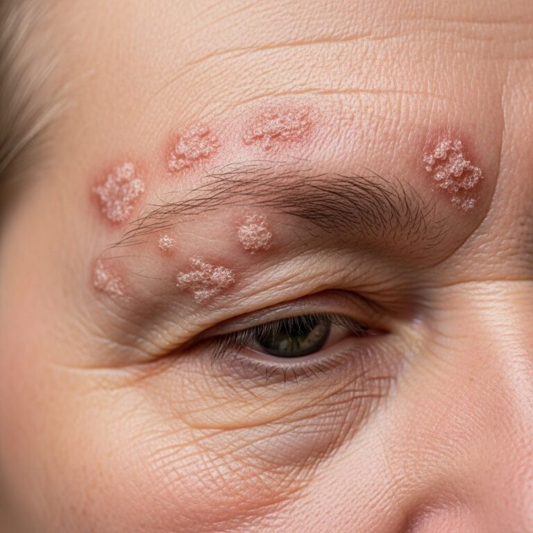
Lymphomatoid papulosis (LyP) is a rare, indolent form of cutaneous T-cell lymphoma characterized by recurrent crops of self-healing papules and nodules on the skin. Despite its malignant histological appearance, LyP behaves benignly, with lesions typically resolving spontaneously within 2–12 weeks, often leaving scars or post-inflammatory pigmentation changes. It represents the most common primary cutaneous CD30+ lymphoproliferative disorder, accounting for approximately 12% of all cutaneous T-cell lymphomas.
Clinically, LyP manifests as erythematous papules or nodules that may ulcerate, crust, or necrose centrally before regressing. The condition waxes and wanes over years or decades, with new crops emerging unpredictably. Although not life-threatening, LyP carries a significant risk of progression to more aggressive lymphomas in 10–25% of cases, including mycosis fungoides, primary cutaneous anaplastic large-cell lymphoma (pcALCL), or Hodgkin lymphoma.
Who gets lymphomatoid papulosis?
LyP affects individuals across all ages, with a mean diagnostic age of 45 years, though pediatric and elderly cases occur. It has a 2:1 male predominance and equal racial distribution, though less frequently reported in darker skin types. Incidence is estimated at 1.2–1.9 cases per million population annually, making it a rare entity. Genetic predisposition is unclear, but clonal T-cell receptor (TCR) gene rearrangements are detected in most lesions, supporting its lymphoproliferative nature.
- Age range: Children to elderly; peak 40–60 years
- Sex: Males > females (M:F ≈ 1.5–2:1)
- Race/ethnicity: All races; underreported in Black skin
- Risk factors: Older age, monoclonal TCR rearrangement increase lymphoma progression risk
Causes
The precise aetiology of LyP remains unknown, but it involves oligoclonal or monoclonal expansion of activated CD30+ T-lymphocytes with malignant potential. These atypical cells resemble Reed-Sternberg-like cells seen in Hodgkin lymphoma. No consistent viral, environmental, or hereditary triggers have been identified, though associations with immunosuppression or prior lymphomas exist.
Histologically, LyP subtypes (A–E, HPV+) are defined by cellular infiltrate patterns: Type A (most common, polymorphous), Type B (mycosis fungoides-like), Type C (monomorphic large cells), Type D (CD8+ cytotoxic), and rarer variants. Pathogenesis likely stems from dysregulated T-cell apoptosis, leading to self-limited proliferations that mimic lymphoma.
Clinical features
Lesions arise in crops of 1–100+ papules (2–20 mm), favouring trunk, extremities, and flexural areas; mucosal, palmoplantar, or generalized involvement is rare. Evolution progresses from pink/red macules to infiltrated papules/nodules, often developing central necrosis, haemorrhagic crusting, or ulceration within days. Mild pruritus affects 20–50% of patients.
Individual lesions involute over 2–8 (up to 12) weeks, leaving varioliform scars, hypopigmentation, or atrophy. Disease duration spans years to decades with intermittent flares; spontaneous complete remission occurs in ~10–20%, though recurrence is common.
| Stage | Duration | Appearance |
|---|---|---|
| Early | Days 1–3 | Erythematous macule/papule |
| Mature | Days 4–10 | Nodule ± haemorrhage/vesiculation |
| Late | Weeks 2–12 | Ulcer/crust → scar |
Complications
Scarring (pitted/atrophic) from necrotic lesions impacts cosmesis, particularly on visible sites. Secondary bacterial infection complicates ulcerated crops. Most critically, 15–25% develop associated lymphomas (cutaneous or systemic), which may precede, coincide, or follow LyP by years. Long-term surveillance is essential.
- Cosmetic: Scarring, dyspigmentation
- Infectious: Cellulitis from ulceration
- Malignant: Mycosis fungoides (40%), pcALCL (20%), Hodgkin (15%), systemic ALCL
Diagnosis
Diagnosis mandates clinicopathological correlation: characteristic history/exam plus biopsy showing CD30+ atypical lymphocytes (≥75% in Type C/D) amid mixed inflammatory infiltrate. Immunohistochemistry (CD3+, CD4+/CD8+ variable, CD30+, CD15−) and TCR gene analysis aid subtyping. Multiple biopsies may be needed to exclude mimics.
Exclusion of extracutaneous disease via bloodwork (CBC, LDH, Sézary count), imaging (CT/PET), or bone marrow is reserved for suspicious features (B-symptoms, lymphadenopathy).
Histological subtypes
- Type A: 75%; pleomorphic CD30+ cells + eosinophils/neutrophils
- Type B: 5–10%; small cerebriform cells (MF-like)
- Type C: 5–10%; sheets of large anaplastic CD30+ cells
- Type D: CD8+ cytotoxic; angioinvasive
- Type E: Epidermotropic, ulceronecrotic
- HPV+: Follicular mucinosis
Differential diagnoses
| Condition | Key Distinguishers |
|---|---|
| Pityriasis lichenoides | More polymorphic; less CD30+; acute/chronic forms |
| pcALCL | Larger solitary lesions; <50% regress |
| Mycosis fungoides | Patches/plaques; persistent |
| Arthropod bites | Grouped; eosinophil-rich; no clonality |
| Viral exanthem | Fever, systemic; self-limited |
Treatment
No curative therapy exists; management targets symptom control, flare reduction, and cosmesis. Observation suits infrequent, asymptomatic crops. Escalation depends on burden, flares, and scarring risk.
First-line (limited disease)
- Topical corticosteroids (potent)
- Intralesional steroids
- Topical nitrogen mustard, carmustine
Second-line (extensive)
- Phototherapy: Narrowband UVB, PUVA
- Low-dose methotrexate (5–35 mg/week)
- Photodynamic therapy
Advanced/refractory
- Oral bexarotene, IFN-α
- Brenuximab vedotin (anti-CD30)
- Low-dose radiotherapy (localized)
Relapse post-discontinuation is universal; treatments do not alter lymphoma risk.
Outcome
Excellent prognosis: 100% 5-year survival, indolent course over decades. ~80% remain LyP-only; 20% develop lymphoma (median 3–5 years post-diagnosis). Annual dermatologic surveillance (exam, biopsy suspicious lesions) is recommended lifelong.
Frequently Asked Questions
Is lymphomatoid papulosis a cancer?
Histologically malignant-appearing but clinically benign; classified as indolent lymphoma with self-resolving lesions.
Will my LyP go away on its own?
Individual crops resolve in 2–12 weeks; disease persists chronically with flares.
Do I need regular check-ups?
Yes, annual skin exams ± biopsy to monitor for lymphoma progression.
Can treatments prevent lymphoma?
No; therapy controls skin symptoms only.
Is LyP contagious?
No, not infectious.
References
- Lymphomatoid papulosis – Patient Information Leaflet — Newcastle Hospitals NHS Foundation Trust. 2023. https://www.newcastle-hospitals.nhs.uk/services/dermatology/patient-dermatology-information-leaflets/lymphomatoid-papulosis/
- Lymphomatoid papulosis — DermNet NZ. 2024-10-15. https://dermnetnz.org/topics/lymphomatoid-papulosis
- Lymphomatoid Papulosis — StatPearls [Internet]. https://www.ncbi.nlm.nih.gov/books/NBK532295/
- Diagnosis: Lymphomatoid Papulosis — Cutaneous Lymphoma Foundation. 2023. https://www.clfoundation.org/lymphomatoid-papulosis
Similar Articles


Read full bio of medha deb