Macular Amyloidosis: Causes, Diagnosis, And Treatment Guide
Understanding the causes, symptoms, diagnosis, and management of macular amyloidosis, a common itchy hyperpigmented skin disorder.
Author: Dr. Purva M. Mehta, Dermatologist
Synonyms: Macular amyloidosis is a form of primary localised cutaneous amyloidosis (PLCA).
What is macular amyloidosis?
**Macular amyloidosis** is the most common form of
primary localised cutaneous amyloidosis
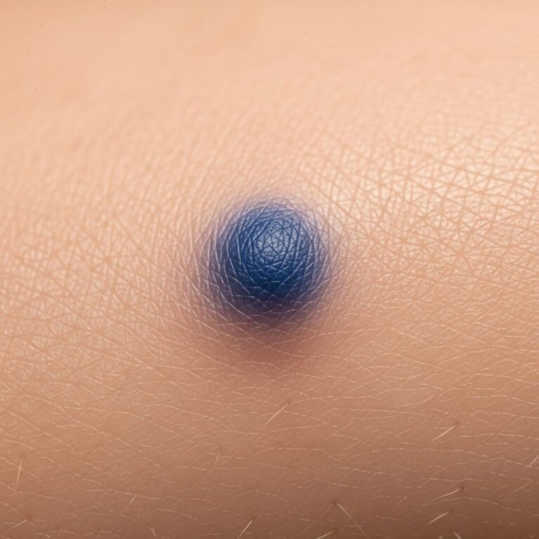
(PLCA). It presents as pruritic macules (flat patches) that are rippled, reticulated, or reticulate-like hyperpigmented lesions measuring 1–10 mm in diameter. These typically appear on the upper back but can also affect the upper arms, chest, abdomen, thigh, or neck. The colour ranges from brown to grey-brown. Itching (pruritus) is the main symptom.Lesions coalesce to form larger confluent patches, resulting in a reticulated (net-like) or mosaic pattern. In chronic cases, the epidermis may thicken (lichenification), and the skin may take on a rippled appearance. Individual lesions are usually smaller than 1 cm in diameter, but they often merge into larger patches.
Macular amyloidosis is more prevalent in Asia, particularly among individuals of Asian descent, with a female predominance. It typically affects adults aged 20–50 years.
Who gets macular amyloidosis?
Macular amyloidosis is most common in Asia, affecting up to 80% of patients presenting with primary localised cutaneous amyloidosis (PLCA). Women are more often affected than men, with a female-to-male ratio of about 2:1.
- Age: Most common in adults aged 20–50 years.
- Race: Predominantly affects individuals of Asian descent, including South Asians, East Asians, and Middle Eastern populations.
- Sex: Female predominance.
What causes macular amyloidosis?
The precise cause of macular amyloidosis is uncertain, although there appears to be an interplay between genetic and environmental factors that is triggered by prolonged periods of friction, rubbing, or scratching of the affected region.
Two main theories explain the aberrant amyloid deposition:
- Fibrillar body theory: Degenerating keratinocytes release tonofilaments that form amyloid fibrils.
- Secretory theory: Local secretion of amyloid precursor proteins by fibroblasts or macrophages.
Risk factors include:
- Chronic friction from bathing practices (e.g., use of loofah, nylon brushes, or bath stones).
- Genetic predisposition, with familial cases reported.
- Possible associations with UV exposure or other irritants.
What are the clinical features of macular amyloidosis?
Macular amyloidosis typically presents with:
- Itchy, hyperpigmented macules (1–10 mm) on the upper back, often in a rippled, reticulated, or ‘crazy-paving’ pattern.
- Lesions may coalesce into larger patches with a net-like or mosaic appearance.
- Skin colour ranges from brown to grey-brown; chronic scratching leads to lichenification (thickened, leathery skin).
- Pruritus is the hallmark symptom, often worse at night.
- Other sites: upper arms, chest, abdomen, thighs, shins, or neck.
In advanced cases, papules may develop (known as biphasic amyloidosis when macular and papular forms coexist). The condition is chronic and persistent, significantly impacting quality of life due to cosmetic concerns and intractable itching.
Diagnosis of macular amyloidosis
Diagnosis is confirmed by skin biopsy showing amyloid deposition in the papillary dermis.
Histology
Key histological features include:
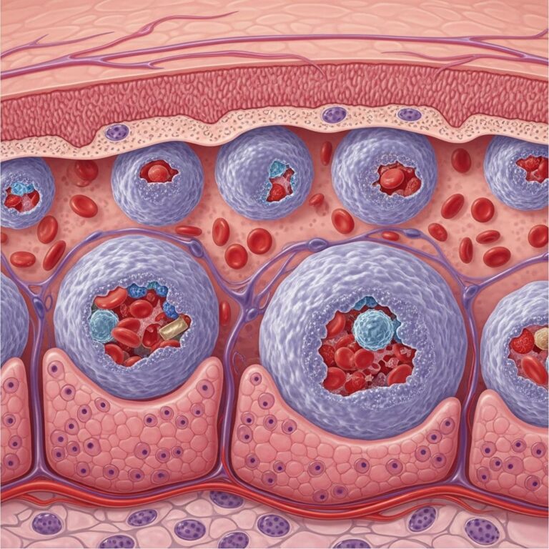
- Amorphous, eosinophilic amyloid deposits in the papillary dermis.
- Perivascular and perifollicular distribution.
- Apple-green birefringence under polarised light after Congo red staining.
Special stains confirm amyloid:
- Congo red: Orangeophilia with apple-green birefringence.
- Thioflavine T: Yellow fluorescence under UV light.
Electron microscopy reveals nonbranching fibrils 7.5–10 nm in diameter.
Related Stories


Differential diagnosis
Macular amyloidosis can be confused with other pigmentary disorders, including:
| Condition | Key Distinguishing Features |
|---|---|
| Notch of hyperpigmentation (friction melanosis) | Larger patches from chronic friction; no amyloid on biopsy. |
| Postinflammatory hyperpigmentation | History of inflammation; resolves over time. |
| Lichen simplex chronicus | Thickened plaques from scratching; no amyloid. |
| Fixed drug eruption | Recurrent at same site; resolves with dusky centre. |
| Photomelanosis (extensor erythema/abrasion folliculitis) | Rippled pattern on extensors; common in Indians; no pruritus. |
What is the treatment for macular amyloidosis?
No standardised treatment regimen has been established, as macular amyloidosis is chronic and not curable. The primary goal of treatment is the relief of pruritus and cosmetic improvement. Emphasis should be placed on avoiding mechanical stressors such as friction, rubbing, or scratching.
Topical treatments (first-line)
- Emollients: To hydrate and protect the skin.
- Topical corticosteroids: Potent agents under occlusion for itch relief.
- Intralesional corticosteroids: For localised thick lesions.
- Topical calcineurin inhibitors: Tacrolimus 0.1% or pimecrolimus 1% ointment, applied for 30 minutes then rinsed.
- Other topicals: DMSO 10%, vitamin D analogues.
Systemic treatments (refractory cases)
- Antihistamines: For pruritus control.
- Retinoids: Acitretin or etretinate (relapse common on discontinuation).
- Immunosuppressants: Cyclosporine, cyclophosphamide (limited efficacy).
Physical therapies
- Phototherapy: Narrowband UVB, broadband UVB, PUVA.
- Laser therapy: Q-switched Nd:YAG (532/1064 nm), pulsed dye laser, fractional lasers, CO2 laser.
- Other procedures: Dermabrasion, cryosurgery, electrodesiccation, TENS.
Treatment success varies; many patients achieve partial improvement in pigmentation and pruritus. IL-31 inhibitors are emerging for pruritus management.
Prevention and lifestyle measures
- Avoid friction: No loofahs, harsh scrubbing, or tight clothing.
- Moisturise regularly to prevent dryness-induced scratching.
- Manage associated atopy or skin conditions.
- Sunscreen on exposed areas to prevent worsening.
Outlook and complications
Macular amyloidosis is benign and confined to the skin (does not progress to systemic amyloidosis). However, it is persistent, with relapses common. Chronic pruritus and cosmetic disfigurement can impair quality of life. Rarely associated with autoimmune diseases in PLCA spectrum.
Frequently asked questions
What causes the itchy brown patches on my upper back?
These are likely macular amyloidosis from amyloid deposits triggered by friction and genetic factors. A biopsy confirms diagnosis.
Is macular amyloidosis curable?
No, but symptoms can be managed with topicals, phototherapy, and avoiding friction. No standardised cure exists.
Does it spread to internal organs?
No, macular amyloidosis is localised to skin only.
How do I stop the itching?
Use emollients, topical steroids/calcineurin inhibitors, and antihistamines. Avoid scratching.
Can lasers remove the pigmentation?
Q-switched Nd:YAG lasers show promising results for pigmentation reduction.
Is it common in Indians?
Yes, prevalent in Asian skin types due to friction from bathing practices.
References
- What is Macular Amyloidosis and How Is It Treated? — BuzzRx. 2023. https://www.buzzrx.com/blog/what-is-macular-amyloidosis-and-how-is-it-treated
- An Interesting Case of Macular Amyloidosis With No Significant History of Friction. — PMC (NCBI). 2024-04-01. https://pmc.ncbi.nlm.nih.gov/articles/PMC11016991/
- Macular Amyloidosis: Causes, Diagnosis and Treatment. — MFine. 2023. https://www.mfine.co/guides/macular-amyloidosis-indians
- Macular amyloidosis. — DermNet NZ. 2024. https://dermnetnz.org/topics/macular-amyloidosis
- Macular Amyloidosis: A Comprehensive Guide. — Dr. Alpana. 2023. https://www.dralpana.com/post/macular-amyloidosis
Similar Articles





Read full bio of medha deb