Macular Degeneration Diagnosis: 9 Key Tests For Early Detection
Learn how macular degeneration is diagnosed, from eye exams to advanced dark adaptometry tests like AdaptDx.

Age-related macular degeneration (AMD) is the leading cause of vision loss in people over 50 in developed countries. Early diagnosis is crucial because while there’s no cure, treatments can slow progression and preserve vision. Diagnosing AMD involves a combination of patient history, symptom evaluation, and specialized eye tests.
What Is Macular Degeneration?
**Age-related macular degeneration (AMD)** affects the macula, the central part of the retina responsible for sharp, detailed vision needed for reading, driving, and recognizing faces. There are two main types: dry AMD (about 80-90% of cases), which involves gradual thinning of the macula, and wet AMD, where abnormal blood vessels leak fluid, causing rapid damage.
Symptoms include blurred central vision, distorted lines (straight lines appear wavy), dark or blank spots in vision, and difficulty adapting to low light (night blindness). Risk factors encompass age over 50, family history, smoking, high blood pressure, obesity, and light-colored eyes.
Who Is at Risk for Macular Degeneration?
Anyone over age 50 should be vigilant, especially with risk factors. According to the National Eye Institute, about 11 million Americans have some form of AMD, with prevalence rising sharply after 60. Early detection is key since damage is irreversible, but interventions like AREDS2 supplements for dry AMD or anti-VEGF injections for wet AMD can help.
- Age: Primary risk; incidence doubles every decade after 50.
- Genetics: Family history increases odds by 2-3 times.
- Lifestyle: Smoking doubles risk; obesity and poor diet contribute.
- Other: Cardiovascular disease, prolonged sun exposure without UV protection.
Macular Degeneration Symptoms to Watch For
AMD often develops silently in early stages. Watch for:
- Blurry or fuzzy central vision.
- Difficulty reading or seeing fine details.
- Wavy or distorted central vision (metamorphopsia).
- Need for brighter light to see objects.
- Colors appearing less vibrant (faded).
- Dark, blurry, or blank spots in center of vision (scotoma).
- Slower night vision adaptation.
If you notice these, especially after 50, schedule a comprehensive dilated eye exam immediately.
Comprehensive Dilated Eye Exam
The cornerstone of AMD diagnosis is a
comprehensive dilated eye exam
. Your eye doctor uses drops to widen pupils, allowing a detailed view of the retina. They look for:- Drusen (yellow deposits under retina) – hallmark of dry AMD.
- Pigment changes in macula.
- Geographic atrophy (late dry AMD).
- Neovascularization or fluid (wet AMD signs).
This painless exam takes 30-60 minutes and is recommended annually for those over 50 or at risk.
Amsler Grid Test
A simple self-test using an
Amsler grid
– a square of parallel lines with a central dot. Cover one eye, stare at the dot, and note distortions or missing lines. Wavy, faded, or missing lines in one or both eyes suggest AMD. Use monthly at home, but it’s not diagnostic – follow up with a doctor.How to use:
- Remove glasses or wear reading ones.
- Hold grid 14-16 inches away at eye level.
- Test each eye separately.
- Normal: All lines straight, uniform; no distortions.
Optical Coherence Tomography (OCT)
**OCT** is a non-invasive imaging test using light waves to create cross-sectional retina images (like an optical ultrasound). It detects fluid, thinning, or drusen invisible during standard exams. OCT angiography (OCTA) visualizes blood flow without dye injection, identifying wet AMD vessels precisely.
Highly sensitive for early changes; repeat scans monitor progression.
Fundus Photography and Fluorescein Angiography
Fundus photography captures detailed retina color photos for baseline and comparison. Fluorescein angiography involves dye injection; serial photos show leakage or abnormal vessels confirming wet AMD. It’s more invasive but definitive when OCT is inconclusive.
Dark Adaptometry: The AdaptDx Test
One of the most promising tools for
early AMD detection
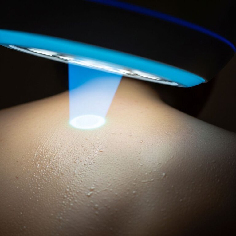
is dark adaptometry**, measuring rod photoreceptor recovery time after bright light exposure. AMD impairs this ‘dark adaptation,’ causing night blindness – an early symptom.The
AdaptDx Pro
from Maculogix automates this. Patient fixates on a target in a dark field; a green flash bleaches rods, then dim lights test recovery time. Normal: 6.5 minutes; prolonged (>6.5 min) signals AMD risk up to 3 years before visible damage.- Screener mode: 5-7 minutes during routine exams.
- Full test: 15-20 minutes for confirmation.
Studies from Harvard, Penn State, Johns Hopkins show 90% sensitivity/specificity for AMD detection. It’s objective, reproducible, and detects preclinical disease, enabling early interventions like quitting smoking, diet changes, or monitoring.
Other Diagnostic Tests
- Fundus Autofluorescence (FAF): Detects lipofuscin in retinal cells, indicating stressed RPE.
- Visual Field Testing: Maps scotoma extent.
- Low-Luminance Visual Acuity: Tests vision in dim light, correlating with progression risk.
How Is Macular Degeneration Staged?
AMD stages:
| Stage | Description | Key Features |
|---|---|---|
| Early | Medium drusen (>63µm), no/minimal symptoms | Monitor; lifestyle advice |
| Intermediate | Large drusen (>125µm), pigment changes, mild vision loss | AREDS2 supplements |
| Late Dry (Geographic Atrophy) | RPE/macula cell loss | Syfovre/Syolntamab injections |
| Late Wet | Neovascularization, rapid loss | Anti-VEGF injections |
Differential Diagnosis
AMD mimics include diabetic retinopathy, central serous chorioretinopathy, histoplasmosis, and myopic degeneration. Tests distinguish them via imaging patterns.
What Happens After Diagnosis?
Treatment depends on type/stage:
- Dry: Lifestyle (quit smoking, leafy greens, exercise), AREDS2 vitamins.
- Wet: Injections (Avastin, Eylea), photodynamic therapy.
- Advanced: Low-vision aids, rehab.
Regular monitoring prevents progression.
Prevention and Early Detection Tips
- Annual dilated exams after 50.
- Home Amsler grid.
- UV-protective sunglasses.
- Healthy diet (omega-3s, antioxidants).
- No smoking; manage BP/cholesterol.
Frequently Asked Questions (FAQs)
Can macular degeneration be diagnosed at home?
No, but the Amsler grid screens for changes. See an eye doctor for confirmation.
How early can AdaptDx detect AMD?
Up to 3 years before traditional signs, by measuring impaired dark adaptation.
Is the AdaptDx test painful?
No, it’s non-invasive, like staring at a screen in a dark room for 5-20 minutes.
Who should get dark adaptometry?
Adults over 50, especially with risk factors or night vision complaints.
Can glasses fix AMD vision loss?
No, AMD affects the retina; glasses aid refraction but not macular damage.
How often should high-risk patients be tested?
Every 6-12 months, or as doctor-recommended based on progression.
References
- Dark Adaptometry with the AdaptDx Pro — Doctor Eye Health (Dr. Joseph J. Allen, OD FAAO). 2021-08-16. https://www.youtube.com/watch?v=mR6d4QjN6KE
- Dark adaptometry as a biomarker for age-related macular degeneration — National Center for Biotechnology Information (PMC). 2014. https://www.ncbi.nlm.nih.gov/pmc/articles/PMC3954002/
- AdaptDx dark adaptometry in AMD — National Center for Biotechnology Information (PMC). 2016. https://www.ncbi.nlm.nih.gov/pmc/articles/PMC4724453/
- Age-Related Macular Degeneration — National Eye Institute (NEI.nih.gov). 2023. https://www.nei.nih.gov/learn-about-eye-health/eye-conditions-and-diseases/age-related-macular-degeneration
- AMD Diagnosis Guidelines — American Academy of Ophthalmology. 2024-05-15. https://www.aao.org/eye-health/diseases/amd-diagnosis
Similar Articles


Read full bio of Sneha Tete