Magnetic Resonance Angiography (MRA): What To Expect
Non-invasive vascular imaging using magnetic fields to visualize blood vessels and detect abnormalities.
: What To Expect")
What is Magnetic Resonance Angiography (MRA)?
Magnetic resonance angiography (MRA) is a specialized imaging technique that uses a powerful magnetic field, radio waves, and computer technology to create detailed images of blood vessels throughout the body. Unlike traditional angiography, which requires inserting a catheter into the body, MRA is a non-invasive procedure that allows physicians to evaluate blood flow and identify vascular abnormalities without the risks associated with catheter insertion. The procedure provides exceptional visualization of both arteries and veins, making it an invaluable diagnostic tool in modern medicine.
MRA is a type of magnetic resonance imaging (MRI) that specifically focuses on the vasculature rather than the surrounding tissues. This targeted approach enables radiologists to obtain high-resolution images of blood vessels in virtually any part of the body, from the brain and neck to the abdomen and extremities. The technique does not expose patients to ionizing radiation, making it a safer alternative to CT angiography for many patient populations, including pregnant women and those requiring multiple imaging studies.
How Does MRA Work?
Unlike X-ray and computed tomography (CT) exams that utilize radiation, MRA functions through a fundamentally different mechanism. The procedure works by using a powerful magnetic field that aligns hydrogen atoms naturally present within the body. As the hydrogen atoms return to their normal alignment after the magnetic field is removed, they emit varying amounts of energy depending on the tissue type. The MRI scanner captures these energy emissions and uses computer processing to convert them into detailed images.
In most MRI units, the magnetic field is produced by passing an electric current through wire coils. Additional coils positioned inside the machine and sometimes placed around the area being imaged send and receive radio waves that produce signals detected by the scanner. Importantly, the electric current does not come into contact with the patient, making the procedure safe for most individuals. A sophisticated computer then processes these signals and creates a series of images, with each image representing a thin slice of the body. Radiologists can examine these images from multiple angles and in three dimensions to obtain comprehensive vascular information.
MRA is often superior to X-ray, CT, and ultrasound in distinguishing between diseased tissue and normal tissue. When contrast material is used, it clearly defines blood vessels by making them appear bright white, enhancing diagnostic accuracy. The contrast material used for MRA is gadolinium-based and is less likely to cause allergic reactions compared to the iodine-based contrast material used in CT angiography.
Clinical Applications of MRA
MRA has extensive applications across numerous vascular conditions and anatomical regions. Healthcare providers utilize this imaging modality for both diagnostic and treatment planning purposes.
Thoracic and Abdominal Conditions
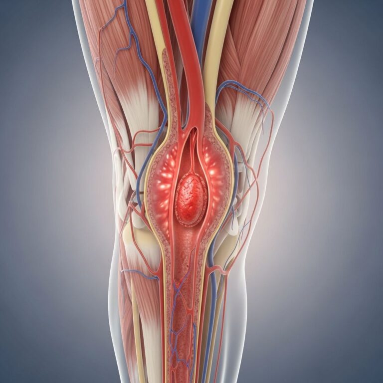
MRA excels at identifying abnormalities in the aorta and its branches throughout the thorax and abdomen. The procedure effectively detects aortic aneurysms, aortic dissections, and stenotic or occlusive vascular disease. Studies demonstrate that MRA has high diagnostic accuracy for pre-operative and post-operative evaluation of aortic dissection and aneurysm, making it comparable to conventional angiography in determining the extent of abdominal aortic aneurysm (AAA). Additionally, MRA can visualize renal artery pathology and blood flow to the kidneys, assisting in surgical planning for AAA repair and kidney transplant evaluation.
Cerebrovascular Applications
In the head and neck region, MRA plays a critical role in stroke prevention and diagnosis. The procedure effectively detects atherosclerotic (plaque) disease in the carotid arteries of the neck, which can limit blood flow to the brain and precipitate stroke. MRA also identifies arteriovenous malformations (AVMs) within the brain and elsewhere, as well as arterial dissections that may result from trauma or other causes.
Peripheral Vascular Disease
For patients with suspected peripheral arterial occlusive disease in the lower extremities, MRA demonstrates superior capability in identifying distal run-off vessels and is competitive with conventional angiography in identifying appropriate inflow vessels. The procedure helps detect plaque disease that has narrowed arteries to the legs and assists in surgical planning for angioplasty/stent placement or surgical intervention.
Pulmonary and Cardiac Applications
MRA can examine pulmonary arteries in the lungs to detect pulmonary embolism (blood clots traveling from leg veins) or pulmonary arteriovenous malformations. In cardiac imaging, MRA demonstrates the extent and severity of coronary artery disease and its effects, facilitating planning for interventions such as coronary bypass and stenting. The technique also provides invaluable evaluation for patients with congenital heart disease, revealing abnormalities in blood vessels and cardiac structures, particularly in pediatric patients.
Additional Clinical Uses
MRA serves multiple other purposes in clinical practice. The procedure guides interventional radiologists and surgeons in making repairs to diseased blood vessels, such as implanting stents or evaluating stent positioning after implantation. It detects arterial injuries sustained during trauma throughout the neck, chest, abdomen, pelvis, and extremities. MRA also evaluates arteries feeding tumors prior to surgery or other procedures such as chemoembolization or selective internal radiation therapy. Furthermore, the procedure screens individuals for arterial disease, particularly those with a family history of vascular disorders.
How is MRA Performed?
Pre-Procedure Preparation
MRA examination is typically conducted on an outpatient basis, though some patients may undergo the procedure during a hospital stay. Patients should arrive with comfortable, loose-fitting clothing and inform their healthcare provider of any metallic implants, pacemakers, or other devices that may interact with the magnetic field. Prior to the examination, patients will be asked to remove clothing, jewelry, watches, hearing aids, and other metallic objects that may interfere with imaging quality.
Procedure Steps
The technologist will position the patient on a moveable exam table, using straps and bolsters to maintain proper positioning and minimize motion artifact. For imaging of specific body regions, devices containing coils capable of sending and receiving radio waves may be placed around or adjacent to the area under examination. These coils enhance signal reception and improve image quality.
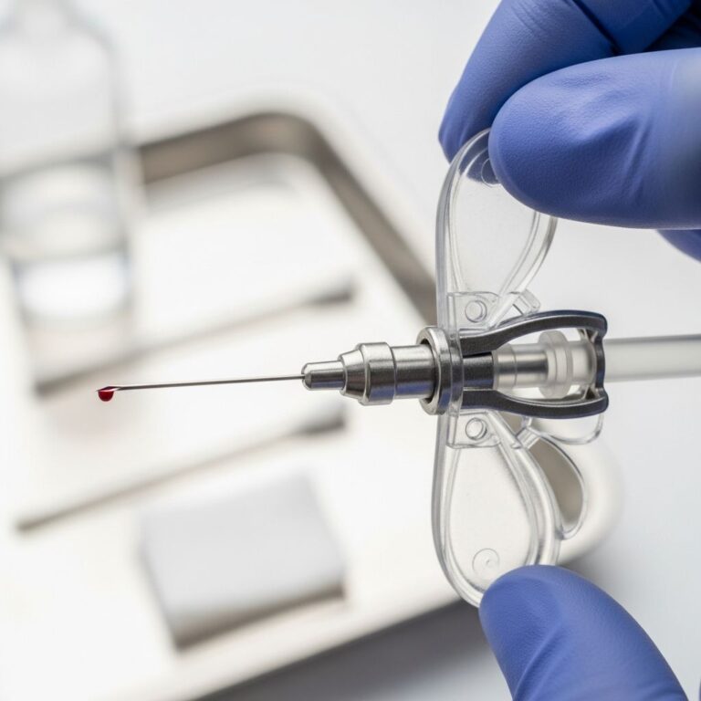
If the MRA requires contrast material, a doctor, nurse, or technologist will insert an intravenous catheter (IV line) into a vein in the patient’s hand or arm before the procedure begins. The patient is then positioned into the magnet of the MRI unit, which resembles a large cylindrical chamber. The technologist operates the equipment from outside the scanning room, and communication occurs through an intercom system, allowing patients to alert staff if they experience discomfort or anxiety.
MRI exams generally include multiple runs or sequences, some of which may last several minutes. Each run produces different acoustic signals, which can sound like knocking, banging, or beeping noises. These sounds are completely normal and do not indicate a problem. If contrast material is being used, the technologist will inject it into the intravenous line after an initial series of scans. Additional images are then acquired during or immediately following the injection to capture the contrast material as it flows through the blood vessels.
Duration and Post-Procedure Care
The entire examination is typically completed in approximately 60 minutes once imaging has started. Upon completion, the technologist may ask the patient to wait briefly while the radiologist reviews the images to determine if additional sequences are needed. The IV line is then removed and a small dressing is placed over the insertion site. Patients may resume normal activities immediately following the procedure.
Safety and Contraindications
Advantages of MRA
MRA offers numerous advantages compared to alternative imaging modalities. The procedure does not utilize ionizing radiation, eliminating cumulative radiation exposure risks associated with repeated imaging. It is a non-invasive technique that avoids the complications inherent to catheter-based angiography, including arterial puncture, thrombosis, and contrast-induced nephropathy. Many MRA protocols can be performed without gadolinium contrast material, benefiting patients with renal insufficiency who require careful consideration regarding gadolinium administration.
Potential Risks and Considerations
While MRA is generally safe, certain precautions must be observed. Patients with metallic implants, including pacemakers, implantable cardioverter-defibrillators (ICDs), ferromagnetic aneurysm clips, or metallic foreign bodies in the eyes should inform their healthcare provider before the procedure. The strong magnetic field can displace or malfunction these devices. Patients may experience claustrophobia within the narrow MRI bore, though open MRI systems are available for anxious patients.
Gadolinium contrast material is generally well-tolerated with fewer allergic reactions than iodine-based contrast. However, in patients with severe renal impairment (estimated glomerular filtration rate less than 30 mL/min/1.73m²), gadolinium administration carries a theoretical risk of nephrogenic systemic fibrosis (NSF), though this risk is minimal with current gadolinium formulations used clinically.
Interpreting MRA Results
After the procedure, a radiologist analyzes the MRA images and generates a detailed report describing the findings. The report will document the presence or absence of vascular abnormalities, including stenosis (narrowing), occlusion (blockage), aneurysms, dissections, and other pathology. The report includes measurements and descriptions of lesion severity and location. Your healthcare provider will discuss the results with you and explain their clinical significance and implications for further management.
Comparison with Other Imaging Modalities
| Imaging Modality | Radiation Exposure | Invasiveness | Contrast Reaction Risk | Cost |
|---|---|---|---|---|
| MRA | None | Non-invasive | Low (gadolinium) | Moderate-High |
| CT Angiography | Yes (significant) | Non-invasive | Moderate (iodine) | Moderate |
| Conventional Angiography | Yes (moderate) | Invasive (catheter) | Moderate (iodine) | High |
| Ultrasound | None | Non-invasive | None | Low |
Frequently Asked Questions
Q: Is MRA painful?
A: No, MRA is not painful. The procedure is non-invasive, though some patients may experience discomfort from lying still or mild sensations from the IV catheter insertion. The acoustic sounds produced by the MRI scanner can be loud, but protective ear plugs are provided.
Q: How long does an MRA take?
A: The entire MRA examination typically takes approximately 60 minutes once imaging has commenced. This includes positioning, potential contrast injection, and multiple imaging sequences.
Q: Can I eat before an MRA?
A: In most cases, there are no dietary restrictions for MRA. However, if the procedure requires IV contrast administration, your healthcare provider may recommend fasting for several hours beforehand as a precaution.
Q: Will I receive contrast material during MRA?
A: Whether contrast material is used depends on the specific clinical question and anatomical region being evaluated. Your healthcare provider will discuss this with you. If used, gadolinium contrast is injected through an IV and enhances visualization of blood vessels.
Q: Is MRA safe for pregnant women?
A: MRA without gadolinium contrast is generally considered safe during pregnancy since it involves no ionizing radiation. However, the use of gadolinium in pregnancy should be carefully discussed with your healthcare provider, as gadolinium does cross the placenta.
Q: What should I wear during the MRA?
A: Wear comfortable, loose-fitting clothes without metallic components. You will be asked to remove all metallic objects including jewelry, watches, hearing aids, and belts before entering the scanning room.
Q: Can patients with pacemakers undergo MRA?
A: Patients with older pacemakers should not undergo MRA due to the strong magnetic field. However, some newer MRI-conditional pacemakers are safe for MRA under specific conditions. Always inform your healthcare provider of any implanted devices.
When to Expect Results
A radiologist will review your MRA images immediately after or within 24 hours of the procedure. Your healthcare provider will typically receive the formal report within one to two business days. Your provider will discuss the results with you and explain any findings and recommended next steps for treatment or additional follow-up imaging if necessary.
References
- MR Angiography (MRA) — Radiology Info. Accessed 2025-01-15. https://www.radiologyinfo.org/en/info/angiomr
- Magnetic Resonance Angiography (MRA) and Magnetic Resonance Venography (MRV) — Aetna. Accessed 2025-01-15. https://www.aetna.com/cpb/medical/data/1_99/0094.html
- MR Angiogram — StatPearls, National Center for Biotechnology Information (NCBI), National Institutes of Health (NIH). Accessed 2025-01-15. https://www.ncbi.nlm.nih.gov/books/NBK558984/
- Magnetic Resonance Angiography (MRA) — University of Rochester Medical Center. Accessed 2025-01-15. https://www.urmc.rochester.edu/encyclopedia/content?contenttypeid=135&contentid=14
- Magnetic Resonance Angiography (MRA) — Cleveland Clinic. Accessed 2025-01-15. https://my.clevelandclinic.org/health/diagnostics/24024-mra
- Definition of Magnetic Resonance Angiography — National Cancer Institute Dictionary of Cancer Terms. Accessed 2025-01-15. https://www.cancer.gov/publications/dictionaries/cancer-terms/def/magnetic-resonance-angiography
- Magnetic Resonance Angiography: MedlinePlus Medical Encyclopedia — National Library of Medicine. Accessed 2025-01-15. https://medlineplus.gov/ency/article/007269.htm
Similar Articles


Read full bio of medha deb