Malignant Histiocytoses: Classification, Diagnosis, and Treatment
Understanding malignant histiocytoses: rare cancers affecting histiocytes with systemic implications and treatment approaches.

Understanding Malignant Histiocytoses
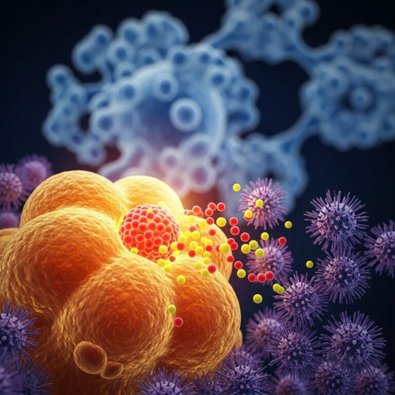
Malignant histiocytoses are cancerous conditions characterized by uncontrolled proliferation of histiocyte cells throughout the body. Also known as Class III histiocytoses, these rare neoplasms represent some of the most aggressive forms of histiocytic disease. Histiocytes are specialized immune cells derived from bone marrow that normally help protect the body by engulfing pathogens and debris. When these cells become malignant, they proliferate uncontrollably and infiltrate various organs, leading to systemic disease with potentially fatal consequences.
The condition typically pursues an aggressive course that may lead to death within a few weeks to months after symptom onset, making early diagnosis and treatment critical. Unlike benign histiocytic conditions, malignant histiocytoses involve abnormal cells that lack normal growth controls and spread rapidly throughout multiple organ systems.
Classification of Malignant Histiocytoses
Malignant histiocytoses encompass several distinct disease entities, each with unique clinical and pathological characteristics:
- Histiocytic sarcoma — a malignant proliferation of cells showing morphologic and immunophenotypic features similar to mature tissue histiocytes
- Monocytic leukaemia — presenting with abnormalities in blood tests and systemic symptoms
- True histiocytic lymphoma — initially presenting as a localized lesion that may later spread throughout the body
In all these conditions, histiocytes spread throughout the body, usually affecting the liver, spleen, lymph nodes, and bone marrow. The classification system helps clinicians and pathologists distinguish between different histiocytic malignancies, enabling more precise diagnosis and treatment planning.
Organs Affected by Malignant Histiocytoses
Malignant histiocytoses typically exhibit a systemic pattern of organ involvement. The primary organs affected include:
| Organ System | Frequency of Involvement | Clinical Significance |
|---|---|---|
| Liver | Common | Often shows hepatomegaly; central to systemic presentation |
| Spleen | Common | Splenomegaly frequently observed; indicates systemic disease |
| Lymph Nodes | Common | Lymphadenopathy present in majority of cases |
| Bone Marrow | Common | Infiltration leads to cytopenias and blood abnormalities |
| Skin | 10-15% | Uncommon but significant when present; rarely the presenting symptom |
| Other Organs | Variable | May include gut, bone, and other tissues |
While skin involvement occurs in only 10-15% of cases, cutaneous manifestations can provide valuable diagnostic clues when present. Malignant histiocytoses may also affect other organs including the skin, making comprehensive evaluation essential for accurate diagnosis.
Clinical Presentations
Patients with malignant histiocytoses present with diverse clinical manifestations depending on which organs are involved. Common systemic symptoms include:
- Fever and malaise
- Weight loss and fatigue
- Lymphadenopathy (enlarged lymph nodes)
- Hepatosplenomegaly (enlarged liver and spleen)
- Pancytopenia (reduction in all blood cell types)
- Leukocytosis (elevated white blood cell count)
- Easy bruising and bleeding
- Recurrent infections
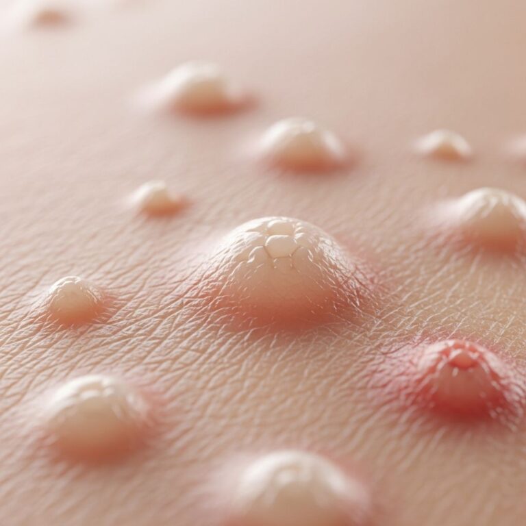
Cutaneous Manifestations
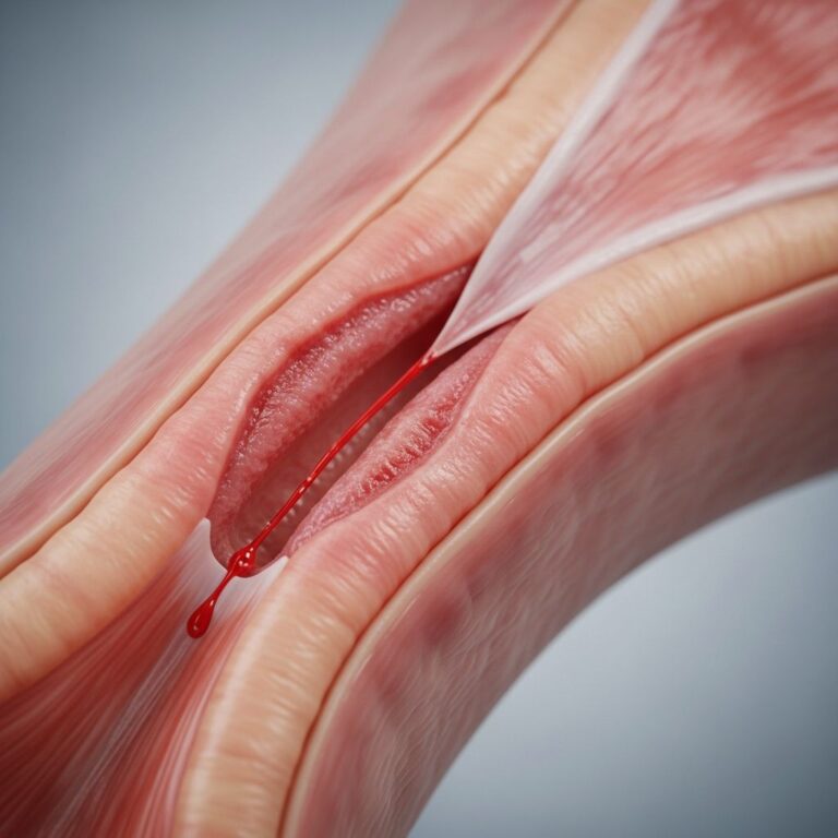
When the skin is involved, lesions typically present as erythematous, maculopapular lesions that can progress to papular or nodular forms. Cutaneous involvement is often polymorphous, making clinical diagnosis challenging. Skin lesions may display purpura, scaling, and/or ulceration, with some patients presenting with violaceous nodules and plaques or morbilliform rashes. The non-healing ulcerated lesions observed in some patients, such as the case of a 74-year-old man with a lesion on his cheek that persisted for several months, demonstrate the aggressive nature of cutaneous involvement.
Specific Disease Presentations
Monocytic leukaemia presents either with unexpected abnormalities found on blood tests or with generalized symptoms of fatigue, weight loss, easy bruising, recurrent infections, skin lumps (leukaemia cutis), and swelling of the gums (gingival hyperplasia). Half of affected people will have involvement of other organs, usually skin, bone, or gut.
True histiocytic lymphoma presents with a localized lump that may be in the lymph glands or in another organ, usually the skin, bone, or gut. It remains localized to one area for some time and may grow quite large before spreading throughout the body.
Histopathological Features
Accurate diagnosis of malignant histiocytoses relies on careful histopathological examination combined with immunohistochemical studies. Key pathological features include:
- Large, pleomorphic epithelioid cells with abundant foamy cytoplasm
- Multilobed nuclei with prominent nucleoli
- Rare erythrophagocytosis (ingestion of red blood cells)
- High mitotic count
- Necrosis often present
- Infiltration into the deep dermis and/or subcutaneous tissues
- Concentration around adnexal structures and blood vessels
- Dyshesive appearance of cells (poor cell-to-cell adhesion)
The presence of large pleomorphic epithelioid cells with foamy cytoplasm, with or without engulfed erythrocytes, should alert dermatopathologists to the possibility of malignant histiocytosis.
Immunohistochemical Staining Profile
Appropriate immunohistochemical evaluation is essential to confirm the diagnosis and distinguish malignant histiocytoses from other conditions. The characteristic immunostaining pattern includes:
Positive Markers:
- CD-68 (diffusely positive in most cases)
- Lysozyme (diffusely positive)
- CD-43 (frequently positive)
Negative Markers:
- CD-1a (negative — important to exclude Langerhans cell histiocytosis)
- S-100 (usually negative — helps exclude dendritic cell disorders)
- CD-3, CD-20, CD-30, CD-34 (negative)
- SMA, MPO (typically negative)
The prominent CD-68 and lysozyme staining along with the histological features and clinical presentation are key diagnostic indicators. Rare examples may show positivity for S-100, CD1a, CD21, CD35, CNA.42, CD34, MPO, CD3, CD20, CD79a, and CD30, though these are uncommon.
Diagnostic Approaches
Malignant histiocytoses are diagnosed through multiple complementary methods:
- Blood tests — reveal cytopenias, leukocytosis, and abnormal cell morphology
- Bone marrow aspirate and biopsy — demonstrates infiltration by malignant histiocytes
- Tissue biopsies — provide histopathological confirmation with immunohistochemical analysis
- Imaging studies — assess organ involvement and disease extent
A comprehensive diagnostic approach combining clinical presentation, laboratory findings, and pathological examination ensures accurate identification and enables appropriate treatment planning. Multiple biopsies from different affected sites may be necessary to confirm systemic involvement, as was demonstrated in the case where six additional random biopsies showed identical histological findings.
Age of Presentation
Malignant histiocytoses can affect patients across a wide age range. Patients afflicted with this condition show ages varying from 2 months to 90 years reported in the medical literature. This broad age spectrum reflects the unpredictable nature of this malignancy and the importance of considering it in differential diagnoses across all age groups.
Treatment Options
Treatment of malignant histiocytoses typically involves aggressive intervention due to the rapid progression of the disease. Most patients with these cancerous forms of histiocytosis respond to treatment with chemotherapy or radiotherapy.
- Systemic chemotherapy — aggressive regimens are usually employed as first-line treatment
- Radiotherapy — may be used for cutaneous lesions or localized disease
- MEK inhibitors — newer agents like trametinib and cobimetinib have demonstrated efficacy regardless of BRAF mutation status
- Local treatments — for cutaneous involvement include topical steroids, calcineurin inhibitors, alkylating agents, phototherapy, steroid injections, and laser therapies
Malignant histiocytosis progresses very quickly, and treatment must be started as early as possible. However, some people will not respond to treatment, and some people will die before the condition can be diagnosed and treated, highlighting the importance of maintaining high clinical suspicion.
Prognosis and Outcomes
Malignant histiocytoses are rare and usually fatal tumors. The condition typically pursues an aggressive course that may lead to death within a few weeks to months after symptom onset. However, recent research suggests that patients presenting with malignant histiocytosis confined to the skin may form a subgroup with a more favorable prognosis. Additionally, spontaneous healing of lesions has been observed, though this is rare and may cause diagnostic confusion.
The variability in outcomes emphasizes the importance of early diagnosis, rapid initiation of treatment, and careful clinical monitoring. Advances in understanding the pathogenesis and development of targeted therapies offer hope for improved outcomes in future patients.
Associated Conditions and Etiopathogenesis
The exact etiopathogenesis of histiocytoses remains poorly understood, though various associations have been identified. Malignant histiocytoses may be associated with:
- Visceral malignancies
- Hematologic cancers
- Autoimmune diseases
- Infections, including Borrelia burgdorferi
These associations are still under active review, and further research is needed to clarify the mechanisms underlying malignant histiocytic transformation and identify potential preventive strategies.
Frequently Asked Questions
Q: What is the difference between malignant histiocytosis and other histiocytic diseases?
A: Malignant histiocytoses (Class III histiocytoses) involve uncontrolled proliferation of abnormal histiocytes with aggressive behavior, in contrast to reactive or benign histiocytic disorders. Malignant forms typically affect multiple organs and pursue a rapidly progressive course.
Q: How common is skin involvement in malignant histiocytoses?
A: Skin involvement occurs in approximately 10-15% of all cases of malignant histiocytosis, making it an uncommon manifestation. However, when present, cutaneous lesions can provide important diagnostic clues.
Q: What immunohistochemical markers are most important for diagnosis?
A: CD-68 and lysozyme are the most important positive markers, showing diffuse staining in malignant histiocytoses. CD-1a and S-100 must be negative to exclude other histiocytic conditions. A comprehensive panel including CD-43, CD-68, CD-3, CD-20, CD-30, CD-34, lysozyme, CD-1a, and S-100 should be used for accurate diagnosis.
Q: Can malignant histiocytosis confined to the skin have a better prognosis?
A: Yes, recent evidence suggests that patients presenting with malignant histiocytosis confined to the skin may form a subgroup with a more favorable prognosis compared to systemic disease, though the condition remains serious and requires prompt treatment.
Q: What is the typical timeline for disease progression?
A: Malignant histiocytosis progresses very quickly, typically leading to death within a few weeks to months after symptom onset. This rapid progression emphasizes the importance of early diagnosis and immediate treatment initiation.
Q: Are newer targeted therapies available for malignant histiocytoses?
A: Yes, MEK inhibitors such as trametinib and cobimetinib have demonstrated efficiency for treating cutaneous histiocytosis, regardless of BRAF mutation status. These represent advances in treatment options beyond traditional chemotherapy and radiotherapy.
References
- Malignant histiocytosis of the skin: a case report and review of the literature — National Center for Biotechnology Information (NIH). 2011-09-15. https://pmc.ncbi.nlm.nih.gov/articles/PMC3157786/
- Clinical and Pathologic Cutaneous Manifestations of Malignant Histiocytosis — JAMA Dermatology. 2012-07-18. https://jamanetwork.com/journals/jamadermatology/fullarticle/544091
- Cutaneous malignant histiocytosis—a clinicopathological review — British Journal of Dermatology. 1985. https://academic.oup.com/bjd/article/113/4/455/6690412
- Unraveling cutaneous histiocytosis: insights into histology, immunophenotyping, and systemic associations — Frontiers in Medicine. 2025-01-15. https://www.frontiersin.org/journals/medicine/articles/10.3389/fmed.2025.1585815/full
- Malignant histiocytoses — DermNet. 2025. https://dermnetnz.org/topics/malignant-histiocytoses
Similar Articles


Read full bio of Sneha Tete