Mastering Silicone Oil Use in Retinal Surgery
Expert techniques for safe silicone oil injection, management, and removal to optimize outcomes in complex vitreoretinal procedures.

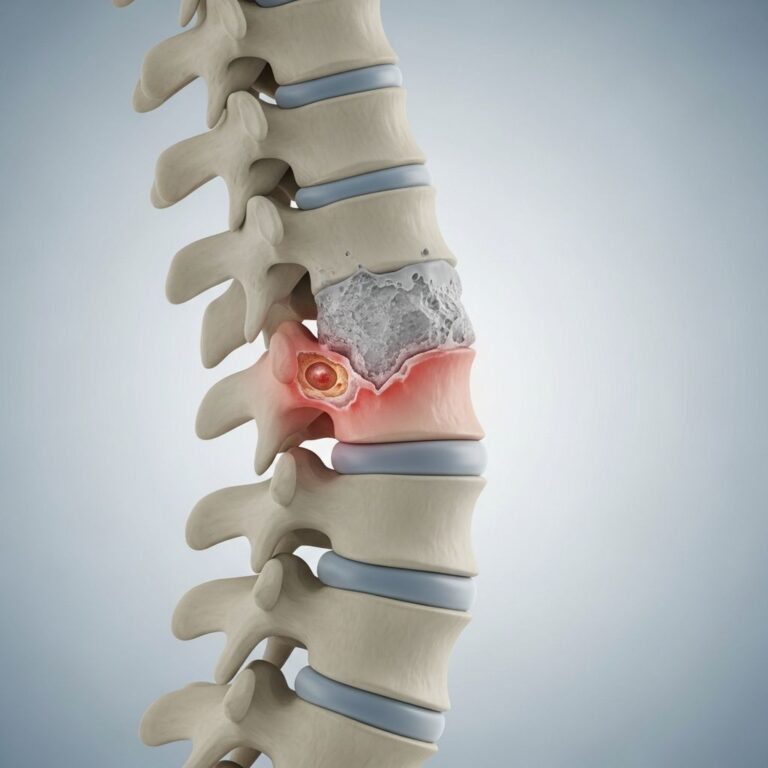
Silicone oil serves as a vital intraocular tamponade agent in vitreoretinal surgery, particularly for managing complex retinal detachments. Its unique physical properties enable it to provide sustained support to the retina, preventing re-detachment in high-risk cases.
Physical Principles Behind Silicone Oil Tamponade
The effectiveness of silicone oil stems from its lower density compared to aqueous and vitreous humor, allowing it to float within the vitreous cavity. This buoyancy generates an upward force, maximal at the apex of the bubble and diminishing toward the horizontal meniscus, creating a tamponade pressure that stabilizes the retina.
Viscosity plays a crucial role in performance. Higher viscosity oils, such as 5000 centistokes (cs), exhibit reduced emulsification tendencies in laboratory settings, prompting many surgeons to select them for long-term use. However, clinical studies reveal no substantial difference in emulsification rates between 1000 cs and 5000 cs oils, influencing material selection based on case-specific needs.
Indications for Silicone Oil in Complex Cases
Surgeons deploy silicone oil primarily in scenarios with elevated redetachment risk, including proliferative vitreoretinopathy (PVR), proliferative diabetic retinopathy (PDR), and viral retinitis-associated detachments. In PDR, it minimizes postoperative hemorrhages and curbs anterior segment neovascularization by blocking vasoproliferative factor migration, thus averting iris rubeosis and neovascular glaucoma.
For PVR cases failing initial vitrectomy, silicone oil offers prolonged tamponade. Decision-making hinges on anticipated tamponade duration, pathology severity, and patient factors like compliance for follow-up oil removal.
Preparation and Equipment for Injection
Modern vitrectomy systems facilitate precise silicone oil delivery via surgeon-controlled foot-pedal pumps connected to syringes. Poiseuille’s law underscores the need for wide-bore syringes, short nondistensible infusion lines, and low-resistance tubing to manage high viscosity and pressures without complications.
- Large syringe: Accommodates elevated infusion pressures.
- Short infusion line: Minimizes flow resistance.
- Nondistensible tubing: Prevents unintended oil injection from elastic recoil.
For 23- or 25-gauge systems, settings limit infusion to 28 psi and aspiration to 30 mmHg to safeguard cannula stability.
Surgical Techniques for Silicone Oil Placement
Three primary methods exist for introducing silicone oil:
- Fluid-silicone exchange: Direct replacement of intraocular fluid with oil using active or passive aspiration.
- Air-silicone exchange: Follows fluid-air exchange; venting cannula positioned posterior to the lens expels air as oil enters, with gradual infusion pressure reduction to 10-12 mmHg for complete fill.
- Perfluorocarbon liquid (PFCL)-silicone exchange: Extrusion needle injects oil while aspirating PFCL, ensuring retinal flattening and superior tamponade.
In air-silicone exchanges, initial high infusion supports intraocular pressure, preventing hypotony. Venting occurs faster for air than oil ingress, necessitating tip placement behind the lens for optimal air evacuation and meniscus visualization.
| Method | Advantages | Considerations |
|---|---|---|
| Fluid-Silicone | Simple, direct | Hypotony risk |
| Air-Silicone | Complete air removal | Pressure management critical |
| PFCL-Silicone | Retinal stabilization | Requires PFCL aspiration |
Anterior Segment Management During Oil Placement
Aphakic patients demand inferior peripheral iridectomy (IPI) to permit aqueous flow beneath the oil bubble, averting pupillary block. This procedure ensures pressure equilibrium and prevents glaucoma.
Phakic eyes require vigilant monitoring to avoid lens contact, which could induce cataract formation. Complete filling targets a visible air meniscus behind the lens, confirming adequate tamponade.
Addressing Emulsification Risks and Prevention
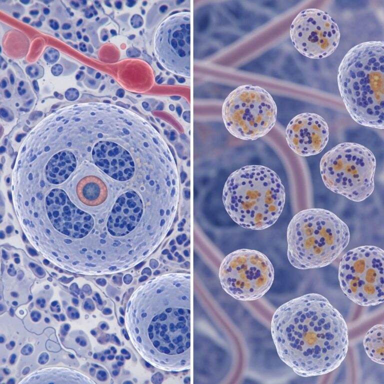
Emulsification represents a primary complication, impacting ocular structures including cornea, trabecular meshwork, retina, and optic nerve. Droplets infiltrate retinal cystoid spaces and optic disc, detectable via optical coherence tomography (OCT).
Duration correlates strongest with emulsification; signs emerge 5-24 months post-injection (average 13 months), universal after 1 year. Routine removal before 12 months is standard unless redetachment risk persists.
- Factors promoting emulsification: Prolonged indwelling time, lower viscosity, inflammation, gas use.
- Prevention strategies: Timely removal, higher viscosity selection, meticulous surgical technique.
Intraretinal oil droplets appear in 21% of PVR cases 3 months post-op via SD-OCT, underscoring early monitoring.
Silicone Oil Removal Procedures
Removal employs specialized sleeves over 23/25-gauge cannulas for aspiration. Tilting removes the sleeve post-evacuation, followed by fluid-air exchanges to clear residual droplets. Anterior chamber emulsified oil necessitates corneal incision evacuation to mitigate keratopathy and glaucoma.
Post-removal, laser retinopexy reinforces attachment. Videos illustrate 25G sutureless techniques in diabetic detachments, highlighting retina flattening during injection.
Complications and Mitigation Strategies
Beyond emulsification, risks include keratopathy, glaucoma, cataracts, and hypotony. IPI mitigates block in aphakics; pressure monitoring prevents over- or under-pressurization.
| Complication | Cause | Mitigation |
|---|---|---|
| Emulsification | Long duration | Remove <12 months |
| Pupillary Block | No IPI in aphakics | Perform IPI |
| Keratopathy | Anterior oil | Clear droplets |
| Glaucoma | Oil in angle | Complete removal |
Advanced Tips for Optimal Outcomes
Pre-injection oil extrusion tests system function. Widefield viewing enhances precision during instillation. In 1000 cs oil cases with 25G systems, superior temporal cannula injection follows air-fluid exchange for combined rhegmatogenous/tractional detachments.
Foot-pedal control allows nuanced pressure adjustments, ensuring 90-100% fill without excess. Postoperative IOP assessment via digital palpation confirms stability.
Patient Selection and Timing
Intra- or preoperative decisions weigh PVR presence, PDR severity, and removal feasibility. Complex cases like failed prior vitrectomies favor oil over gas.
FAQs
What viscosity silicone oil is best?
1000 cs suits most cases; 5000 cs for prolonged tamponade despite similar clinical emulsification.
How long should silicone oil remain?
Typically under 12 months to minimize emulsification; extend if redetachment risk high.
Is IPI always needed?
Mandatory in aphakic oil-filled eyes to prevent block.
What if oil emulsifies anteriorly?
Remove via corneal incision to avoid keratopathy/glaucoma.
Can oil enter the retina?
Yes, OCT shows intraretinal droplets in ~21% PVR cases.
References
- Silicone Oil: Different Physical Properties and Clinical Applications — PMC/NCBI. 2014-06-12. https://pmc.ncbi.nlm.nih.gov/articles/PMC4071776/
- General principles of silicone oil injection. Part 1 — YouTube/RetinaCoach. N/A. https://www.youtube.com/watch?v=qhKFCFaWxeg
- Injection of 1000 Centistokes Silicone Oil using 25G Vitrectomy Technique — Eyetube. 2012-11-15. https://eyetube.net/index.php/videos/injection-of-1000-centistokes-silicone-oil-using-25g-vitrectomy-technique
- Silicone Oil Emulsification in Retina Surgery — Retina Today. 2015-09-01. https://retinatoday.com/articles/2015-sept/silicone-oil-emulsification-in-retina-surgery
- Silicone Oil Extraction Technique — Eyetube. N/A. https://eyetube.net/videos/silicone-oil-extraction-technique
Similar Articles


Read full bio of medha deb