Mastering Single-Pass Four-Throw Pupilloplasty
Discover the innovative SFT pupilloplasty technique revolutionizing iris repair for trauma, glaucoma, and corneal surgeries with minimal invasion.

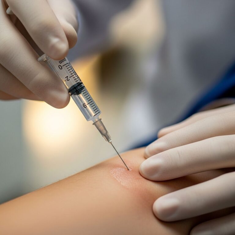
The single-pass four-throw (SFT) pupilloplasty represents a breakthrough in ophthalmic surgery, offering a streamlined approach to reconstructing damaged pupils. This technique uses a single needle pass through the iris followed by four throws around the suture loop to create a self-locking helical knot, minimizing anterior chamber manipulation and reducing complications.
Understanding Iris and Pupil Disorders
The iris, the colored part of the eye, controls light entry through the pupil. Defects from trauma, surgery, or disease distort pupil shape, causing glare, photophobia, and vision impairment. Common issues include irregular pupils post-trauma, iatrogenic damage during cataract surgery, and synechiae in angle-closure glaucoma (ACG), where iris adheres to the cornea or lens, blocking fluid outflow.
In ACG, peripheral anterior synechiae (PAS) exacerbate intraocular pressure (IOP) rise. Traditional repairs often require multiple passes or knots, risking endothelial damage, especially in shallow anterior chambers or post-keratoplasty eyes.
Why Choose SFT Pupilloplasty?
SFT stands out for its simplicity, short learning curve, and versatility. Unlike McCannel or Siepser knots needing two passes, SFT involves one pass, forming a flat, non-bulky helical structure that slides into place without traditional knotting.
- Minimal Invasion: Single pass reduces endothelial trauma and inflammation.
- Self-Locking: Four throws create intertwined loops preventing slippage.
- Versatile Applications: Trauma, cosmetics, Urrets-Zavalia syndrome, pre-Descemet endothelial keratoplasty (PDEK), and ACG.
- Safe for Corneal Grafts: No protruding knots minimize graft-endothelium contact.
Studies show SFT effectively reshapes pupils, breaks PAS, and lowers IOP in ACG by improving aqueous outflow.
Patient Selection and Preoperative Evaluation
Ideal candidates have iris defects >90°, irregular pupils causing symptoms, or ACG with PAS. Exclude active infection, uncontrolled IOP, or severe atrophy where tissue won’t hold sutures.
Preop steps include:
- Slit-lamp exam to measure defect size and synechiae extent.
- Gonioscopy for angle assessment in glaucoma cases.
- Topography and pachymetry for corneal health.
- Patient counseling on risks like IOP spikes, suture erosion (rare).
| Condition | SFT Suitability | Expected Traction Points |
|---|---|---|
| Traumatic Iris Defect | High | 2-4 |
| ACG <270° PAS | High | 4 |
| ACG >270° PAS | High | 6 (3 SFTs) |
| Post-keratoplasty | Excellent (knotless) | 2-4 |
This table outlines traction needs based on synechiae degree, ensuring proportional iris pull.
Step-by-Step Surgical Protocol
SFT demands precision instruments: 10-0 polypropylene on long straight needle, end-opening forceps, Sinskey hook, 30-gauge needle (optional), anterior chamber maintainer (ACM).
- Prepare Access: Mark paracentesis sites 45-90° apart opposite defect. Insert trocar ACM or viscoelastic (if posterior capsule intact). Avoid viscoelastic in aphakia or glued IOL to prevent posterior migration.
- Pupillary Stretch: Use forceps to stretch iris every 1-2 clock hours, breaking synechiae and enhancing elasticity. Critical in ACG.
- First Edge Grasp: Through proximal paracentesis, grasp iris edge with forceps. Pass 10-0 needle through tissue from distal site, leave in situ.
- Loop Creation: Retrieve loop with Sinskey hook via proximal site. Pass suture end through loop four times, forming helical twists.
- Slide and Tighten: Pull both ends evenly; loop slides in, apposing edges. Repeat for multi-point traction if needed.
- Closure: Hydrate chamber, check centration under pupil dilation. No burying required due to flat knot.
Related Stories


Procedure time: 5-15 minutes. Intraop IOP monitoring via ACM prevents spikes.
Advanced Applications in Complex Cases
In endothelial keratoplasty (DMEK/PDEK), SFT’s knotless design prevents donor touch in shallow chambers. For Urrets-Zavalia (fixed dilated pupil post-keratoplasty), SFT constricts and centers pupil, alleviating photophobia.
ACG management: Stretch loosens PAS; SFT sustains traction. For 360° synechiae, three SFTs (6 points) restore angle access, dropping IOP without trabeculectomy.
Cosmetic pupilloplasty for colobomas uses 2-4 SFTs for round pupils, boosting patient satisfaction.
Postoperative Management and Outcomes
Postop regimen:
- Topical steroids/antibiotics taper over 4 weeks.
- Dilation drops initially, then weaning.
- Follow-up: Day 1, Week 1, Month 1, 3, 6 for pupil shape, IOP, endothelium.
Outcomes: 90-95% success in pupil centering, glare reduction. IOP drops 20-30% in ACG. Complications <5%: transient IOP rise, suture cheese-wiring (rare with proper bite).
Long-term: Stable up to 5 years; reoperation rare.
Potential Complications and Mitigation
Though low-risk, anticipate:
| Complication | Incidence | Prevention/Treatment |
|---|---|---|
| IOP Spike | 10% | ACM use; steroids |
| Suture Erosion | <2% | Deep bites; monitor |
| Endothelial Loss | <1% | Single pass; visco if safe |
| Under/Over-Correction | 5% | Preop planning; adjust pulls |
Proactive stretch and ACM minimize risks.
Comparing SFT to Traditional Techniques
SFT excels over Siepser (two passes, bulkier), McCannel (knot burial needed), and Sham (complex loops).
| Technique | Passes | Knot Type | Best For |
|---|---|---|---|
| SFT | 1 | Helical, flat | All, esp. keratoplasty |
| Siepser | 2 | Sliding | Trauma |
| McCannel | 2 | Fixed | Small defects |
SFT’s efficiency suits modern microincision surgery.
Training and Skill Development
Learning curve: 10-20 cases. Wet lab on porcine eyes simulates tissue. Video platforms like EyeTube aid visualization. Proficiency yields repeatable results, expanding armamentarium.
Future Directions
Emerging: Adjustable SFT with laser assistance, bioabsorbable sutures. Research integrates SFT with MIGS for glaucoma combo therapy. AI-guided planning may optimize sites.
Frequently Asked Questions (FAQs)
What is the recovery time after SFT pupilloplasty?
Most resume normal activities in 1-2 weeks; full vision stabilizes by 1 month.
Is SFT suitable for pediatric patients?
Yes, with adjustments for smaller eyes; used in traumatic cases.
Can SFT be combined with cataract surgery?
Absolutely; intraop pupil repair enhances IOL centration.
How does SFT help in glaucoma?
Breaks PAS, opens angles, reduces IOP without shunts.
What suture material is best?
10-0 polypropylene for strength and longevity.
References
- Single-Pass Four-Throw Pupilloplasty — EntoKey. Accessed 2026. https://entokey.com/single-pass-four-throw-pupilloplasty/
- Pupilloplasty — EyeWiki (AAO). Accessed 2026. https://eyewiki.org/Pupilloplasty
- Single-Pass 4-Throw Technique (SFT) For Pupilloplasty — EyeTube (Priya Narang, MS). 2017-05-25. https://eyetube.net/index.php/videos/single-pass-4-throw-technique-sft-for-pupilloplasty
- Single-pass four-throw pupilloplasty for angle-closure glaucoma — PMC (NCBI). 2018. https://pmc.ncbi.nlm.nih.gov/articles/PMC5778544/
- ESCRS Video Of The Month: Single pass 4 throw pupilloplasty — YouTube. 2019. https://www.youtube.com/watch?v=Qa83tQ1-gUw
- Single-pass four-throw technique for pupilloplasty — Ovid (European Journal of Ophthalmology). 2017. https://www.ovid.com/journals/ejoop/fulltext/10.5301/ejo.5000922
Similar Articles


Read full bio of Sneha Tete