Mastocytoma: Essential Guide To Causes, Diagnosis, Treatment
Mastocytoma: A benign skin condition in infants caused by mast cell accumulation, often resolving by puberty.

Mastocytoma is a rare form of
cutaneous mastocytosis
characterised by the accumulation of mast cells in the dermis, forming one to three solitary lesions, most commonly diagnosed in infants aged 0–3 months.What is mastocytoma?
Mastocytoma represents a localised proliferation of mast cells within the skin, classified under cutaneous mastocytosis. Unlike more widespread forms like urticaria pigmentosa, mastocytoma typically presents with 1–3 discrete lesions. Mast cells are immune cells derived from myeloid stem cells, residing in connective tissues such as the skin and mucosa, where they release mediators like histamine upon activation. This release causes localised inflammation, manifesting as redness, swelling, and itching. In mastocytoma, this accumulation is benign and often self-limiting, particularly in childhood-onset cases.
Cutaneous mastocytosis, including mastocytoma, affects children predominantly, with lesions appearing within the first few months of life. It is distinct from systemic mastocytosis, which involves multiple organs and is more common in adults.
Who gets mastocytoma?
Mastocytoma primarily affects infants, with most cases diagnosed between birth and 3 months of age. It is rarely seen in adults, and when it occurs, it warrants investigation for systemic involvement. The condition is not usually familial, though associations with KIT gene mutations have been noted. Cutaneous mastocytosis overall is more common in children, resolving by puberty in many cases, while adult-onset forms like urticaria pigmentosa may persist.
- Peak incidence: 0–3 months of age.
- Rare in adults; consider systemic mastocytosis if present.
- No strong hereditary pattern reported.
Causes
The exact cause of mastocytoma remains unknown but is likely multifactorial. A key genetic factor is a mutation in the
KIT gene
(often c-KIT D816V), which encodes a transmembrane tyrosine kinase receptor essential for mast cell growth, survival, and function. This mutation leads to uncontrolled mast cell proliferation. Activation of these mast cells triggers degranulation, releasing histamine, leukotrienes, prostaglandins, tryptase, and cytokines, causing clinical symptoms.Unlike systemic forms, mastocytoma is confined to the skin and not linked to inheritance. Environmental triggers like friction or temperature changes can provoke symptoms by stimulating mast cell mediator release.
Clinical features
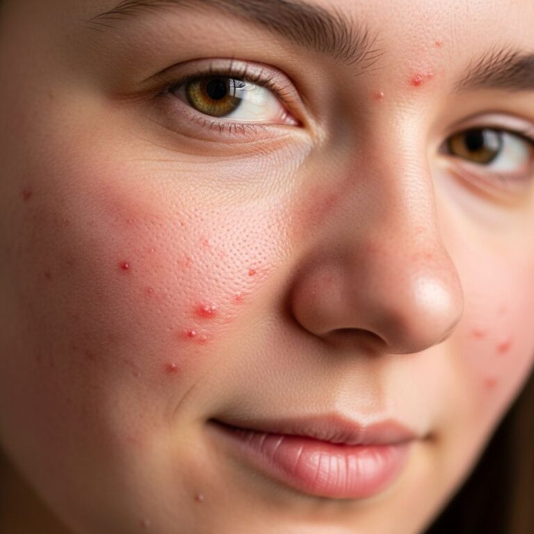
Mastocytoma lesions typically appear as 1–3 solitary, well-circumscribed, reddish-brown or yellowish-brown macules, papules, or plaques, ranging from 1–5 cm in diameter. They can occur on any body site but are often on the trunk or extremities. Key features include:
- Colour: Red-brown, tan, or yellow-brown.
- Shape: Round or oval, raised or flat.
- Symptoms: Pruritus (itching), warmth, and swelling upon stroking (positive
Darier sign
).
Upon mechanical stimulation (rubbing or stroking), lesions exhibit
Darier’s sign
: rapid urtication, erythema, and pseudopod formation due to mast cell degranulation. Occasionally, flushing may occur locally or generalised if chemicals enter circulation. Blistering is rare but possible in infants. In darker skin types (Fitzpatrick IV–VI), lesions may appear hyperpigmented with less erythema, but histology confirms mast cell aggregates.| Feature | Description |
|---|---|
| Lesion Number | 1–3 solitary |
| Size | 1–5 cm |
| Onset | Infancy (<3 months) |
| Darier Sign | Positive: urtication on rubbing |
| Symptoms | Itch, swell, flush |
Severe reactions like anaphylaxis are rare in isolated mastocytoma but possible with extensive rubbing.
Diagnosis
Mastocytoma is often diagnosed clinically, especially with a positive Darier sign in an infant. Dermatoscopy shows non-specific vascular patterns, lacking melanocytic features. Blood tests are usually unnecessary for solitary lesions.
Histology from skin biopsy confirms diagnosis: diffuse or nodular dermal infiltrates of monomorphic mast cells staining positive for
CD117 (c-KIT)
andtryptase
via immunoperoxidase. Aggregates fill the papillary dermis. For adult-onset or symptomatic cases, rule out systemic mastocytosis with serum tryptase, bone marrow biopsy (major criterion: >15% mast cells in clusters), and KIT mutation testing (minor criterion).WHO criteria for systemic mastocytosis include one major and one minor, or three minors.
Differential diagnoses
Mastocytoma may mimic:
- Insect bite reaction: Persistent, itchy nodule.
- Melanocytic naevus: Non-urticating pigmented lesion.
- Urticaria pigmentosa: >4 lesions (maculopapular cutaneous mastocytosis).
- Xanthoma or juvenile xanthogranuloma: Yellowish nodules.
- In adults: Consider telangiectasia macularis eruptiva perstans (TMEP) or systemic disease if Darier positive.
Biopsy distinguishes if clinical doubt exists.
Treatment
Asymptomatic mastocytoma requires no treatment and is managed conservatively. Symptomatic cases focus on avoiding triggers and symptom relief:
- Avoidance: Friction, heat, NSAIDs, opioids, alcohol.
- Topical: Corticosteroids for itch; calcineurin inhibitors (e.g., tacrolimus).
- Oral H1/H2 blockers: Antihistamines (cetirizine, ranitidine) for pruritus/flushing.
- Surgical excision: For solitary lesions in cosmetic areas, though urtication may recur in scars.
For systemic concerns, midostaurin or stem cell transplant in aggressive cases (not typical for mastocytoma). No role for routine mast cell stabilisers like cromolyn in cutaneous forms.
Outcome
Infantile mastocytomas resolve spontaneously before puberty in most cases (>90%). Persistent lesions into adulthood are rare and may indicate indolent systemic mastocytosis (good prognosis, median survival >20 years). Localised mastocytoma has excellent prognosis; mast cell sarcoma (rare extracutaneous) is aggressive.
Monitor for systemic symptoms; annual tryptase if persistent.
Frequently asked questions
Is mastocytoma dangerous?
Solitary mastocytoma in infants is benign and self-resolving. Anaphylaxis risk is low.
Does mastocytoma go away?
Yes, most infantile cases regress by puberty.
What triggers mastocytoma symptoms?
Rubbing, heat, stress provoke Darier sign via degranulation.
Should I biopsy a mastocytoma?
Not routinely for solitary infantile lesions; yes if adult-onset or systemic signs.
Can mastocytoma be treated with medication?
Symptoms respond to antihistamines; excision for select cases.
References
- Mastocytosis (Mast Cell Disorder): Symptoms and Treatment — Patient.info. 2023. https://patient.info/doctor/dermatology/mastocytosis-and-mast-cell-disorders
- Mastocytoma (mast cell birthmark) – DermNet — DermNet NZ. 2023-10-01. https://dermnetnz.org/topics/mastocytoma
- Mastocytosis – DermNet — DermNet NZ. 2023. https://dermnetnz.org/topics/mastocytosis
- Mastocytoma – StatPearls — NCBI Bookshelf (National Center for Biotechnology Information). 2023-07-17. https://www.ncbi.nlm.nih.gov/books/NBK538252/
Similar Articles


Read full bio of Sneha Tete