Medical Imaging: Diagnostic Tests and Therapies
Comprehensive guide to imaging technologies for diagnosis and treatment planning.

Understanding Medical Imaging: Diagnostic Tests and Therapies
Medical imaging plays a crucial role in modern healthcare, enabling physicians to visualize internal structures and functions of the body without invasive surgery. These advanced diagnostic technologies allow doctors to detect disease early, guide treatment decisions, and monitor patient progress over time. From routine screening to complex interventional procedures, imaging modalities have become indispensable tools in clinical practice across virtually every medical specialty.
The field of medical imaging encompasses a diverse range of technologies, each with unique capabilities and applications. Whether evaluating the chest, abdomen, pelvis, brain, or musculoskeletal system, imaging specialists use sophisticated equipment and techniques to create detailed pictures that reveal what is happening inside the body. Understanding these different imaging options helps patients make informed decisions about their healthcare and helps physicians select the most appropriate diagnostic approach for each clinical situation.
Common Medical Imaging Modalities
Modern medicine relies on several primary imaging technologies, each offering distinct advantages for specific clinical applications. These modalities range from traditional radiography to advanced cross-sectional imaging techniques that provide three-dimensional visualization of anatomical structures.
X-Ray Imaging
X-ray imaging represents one of the oldest and most widely available medical imaging technologies. This modality uses electromagnetic radiation to create two-dimensional images of bones, lungs, and other structures. X-rays are particularly valuable for detecting fractures, evaluating lung conditions, and assessing bone density. The technology is fast, inexpensive, and requires minimal radiation exposure, making it an excellent first-line imaging choice for many clinical scenarios. X-ray imaging remains fundamental in emergency departments, primary care settings, and specialized orthopedic practices.
Computed Tomography (CT)
Computed tomography, commonly referred to as CT or CAT scanning, represents a significant advancement in cross-sectional imaging. CT scanners use rotating X-ray beams and computer reconstruction to create detailed three-dimensional images of internal organs and structures. This technology is particularly valuable for evaluating the chest, abdomen, and pelvis, providing exceptional detail for detecting tumors, infections, injuries, and vascular abnormalities. CT imaging is rapid, reproducible, and can be performed with or without intravenous contrast enhancement to optimize visualization of specific structures.
Magnetic Resonance Imaging (MRI)
Magnetic resonance imaging uses powerful magnetic fields and radio frequency waves to generate detailed images of soft tissues, organs, and the nervous system. Unlike CT and X-ray imaging, MRI does not utilize ionizing radiation, making it an excellent choice for imaging the brain, spinal cord, joints, and soft tissues. MRI excels at showing subtle tissue variations and is particularly useful for detecting neurological conditions, musculoskeletal injuries, and abnormalities of the liver and kidneys. Advanced MRI techniques can also provide functional information about brain activity and tissue perfusion.
The physics of MRI involves aligning hydrogen nuclei with a strong magnetic field and then using radio frequency pulses to excite these nuclei. As the nuclei relax and return to their original state, they emit signals that sophisticated computer algorithms reconstruct into detailed images. Different pulse sequences optimize visualization of specific tissues and pathologies, allowing radiologists to tailor the examination to clinical questions.
Ultrasound Imaging
Ultrasound imaging uses high-frequency sound waves to visualize internal structures and is unique among imaging modalities in its lack of ionizing radiation. This real-time imaging technology is ideal for evaluating pregnancy, assessing blood flow in vessels, and examining organs in the abdomen and pelvis. Ultrasound offers several advantages including portability, real-time visualization, and the ability to guide interventional procedures. The versatility of ultrasound extends far beyond obstetric applications, with specialized applications in cardiac imaging, vascular assessment, and musculoskeletal evaluation.
Nuclear Medicine and Molecular Imaging
Nuclear medicine technology represents a cutting-edge field that uses radiopharmaceuticals to visualize molecular and anatomical information. This imaging approach involves administering small amounts of radioactive substances that accumulate in specific tissues or organs, allowing visualization of both structure and function. Nuclear medicine imaging is particularly valuable for evaluating bone metastases, thyroid disorders, cardiac viability, and detecting infections. Positron emission tomography (PET) scanning, a type of nuclear medicine imaging, provides exceptional sensitivity for detecting cancer and evaluating neurodegenerative diseases.
Specialized Imaging Applications
Beyond conventional imaging modalities, Johns Hopkins and other leading medical centers employ specialized imaging techniques for specific clinical purposes. These advanced applications combine traditional imaging technologies with innovative computational methods and interventional capabilities.
Advanced Biophotonics
Novel optical imaging technologies, including fluorescence microscopy and tomographic endoscopy, enable early disease detection at the cellular and molecular level. These techniques provide unprecedented visualization of tissue structure and function, supporting research and clinical applications in oncology, gastroenterology, and other specialties.
Image-Guided Interventions
Modern imaging platforms integrate real-time visualization with interventional capabilities, allowing physicians to perform high-precision procedures with enhanced accuracy. Cone-beam CT, image registration, navigation systems, and robotic assistance enable minimally invasive approaches to biopsy, ablation, and other therapeutic interventions. These image-guided techniques reduce complications, improve outcomes, and decrease patient recovery time compared to traditional surgical approaches.
Advanced Imaging Algorithms
Sophisticated computational methods, including machine learning and high-fidelity physical models, enhance image reconstruction and analysis across all modalities. These algorithms improve image quality, reduce radiation dose in CT and nuclear medicine studies, accelerate image acquisition, and extract quantitative information that supports diagnosis and treatment planning.
Clinical Applications by Organ System
Different imaging modalities excel in evaluating specific organ systems and clinical conditions. Johns Hopkins radiologists employ subspecialty expertise to optimize imaging protocols for each clinical scenario.
Chest Imaging
Chest radiography and CT imaging are essential for evaluating lung diseases, mediastinal masses, and cardiac abnormalities. High-resolution CT provides detailed visualization of lung parenchyma for detecting early interstitial lung disease and characterizing nodules. Cardiac imaging combines CT and MRI with specialized protocols for coronary artery assessment and evaluation of cardiac function.
Abdominal and Pelvic Imaging
Cross-sectional imaging with CT and MRI provides comprehensive evaluation of abdominal and pelvic organs including the liver, pancreas, kidneys, and gastrointestinal tract. Ultrasound offers real-time imaging for emergency evaluation and guidance of interventional procedures. Imaging protocols are tailored to specific clinical questions, such as tumor staging, detection of appendicitis, or evaluation of abdominal pain.
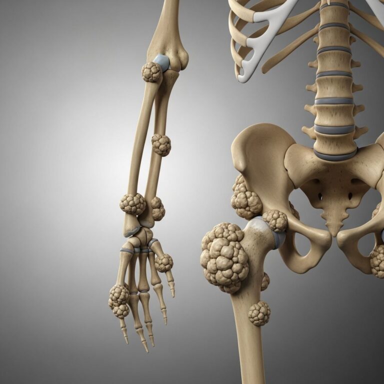
Musculoskeletal Imaging
MRI and ultrasound excel in evaluating joints, muscles, tendons, and ligaments. These modalities provide superior soft tissue contrast for detecting injuries, degenerative changes, and inflammatory conditions. Radiography remains valuable for initial bone assessment and follow-up after orthopedic procedures.
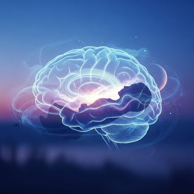
Neurological Imaging
Brain and spinal cord imaging relies primarily on MRI, which provides exceptional detail of gray matter, white matter, and cerebrospinal fluid. Advanced techniques such as diffusion imaging, perfusion imaging, and functional MRI support evaluation of stroke, dementia, multiple sclerosis, and tumor characterization. Image analysis and registration techniques help quantify changes in neurological diseases like Alzheimer’s over time.
Vascular Imaging
CT angiography and MR angiography provide non-invasive assessment of blood vessels throughout the body. Ultrasound with Doppler capability evaluates blood flow characteristics and detects stenosis or aneurysms. These imaging approaches guide treatment decisions in stroke, peripheral arterial disease, and aortic conditions.
Imaging Technology and Education
Leading academic medical centers like Johns Hopkins maintain rigorous educational programs that combine theoretical knowledge with practical clinical experience. Students and residents learn the physics of imaging technologies, device design principles, clinical applications, and advanced computational techniques. Hands-on experience in clinical settings, research laboratories, and classroom environments prepares imaging professionals to advance the field while delivering excellent patient care.
Professional societies and medical centers offer continuing medical education programs to keep practicing radiologists current with rapidly evolving technology and best practices. Johns Hopkins conducts multiple CME courses nationally and internally covering MRI, CT, ultrasound, and emerging imaging techniques. These educational initiatives ensure that imaging specialists maintain expertise and incorporate evidence-based approaches into clinical practice.
Advantages and Considerations of Different Modalities
| Imaging Modality | Primary Advantages | Limitations | Common Clinical Uses |
|---|---|---|---|
| X-Ray | Fast, inexpensive, widely available, low radiation dose | Limited soft tissue contrast, two-dimensional | Fractures, lung evaluation, bone assessment |
| CT | Rapid, excellent detail, three-dimensional, good for trauma | Radiation exposure, less soft tissue contrast than MRI | Tumor staging, trauma evaluation, pulmonary embolism |
| MRI | No radiation, superior soft tissue contrast, functional capabilities | Slower scan time, contraindicated with certain implants, higher cost | Brain imaging, joint evaluation, liver assessment |
| Ultrasound | No radiation, real-time imaging, portable, inexpensive | Operator dependent, limited in obese patients, poor for bone | Obstetrics, vascular assessment, abdominal evaluation |
| Nuclear Medicine | Functional information, high sensitivity for specific pathology | Lower spatial resolution, radiation exposure, longer acquisition time | Cancer detection, bone metastases, cardiac viability |
The Future of Medical Imaging
Medical imaging continues to evolve with advances in hardware, software, and artificial intelligence. Emerging technologies promise improved image quality, faster acquisitions, lower radiation doses, and enhanced quantitative analysis. Machine learning algorithms increasingly assist radiologists in detecting abnormalities, reducing interpretation variability, and predicting clinical outcomes. Integration of imaging data with genomic information and electronic health records will enable increasingly personalized approaches to diagnosis and treatment planning.
Research in imaging physics and engineering focuses on developing novel imaging systems, advanced reconstruction algorithms, and innovative applications of existing technologies. Multi-institutional collaborations and training programs ensure that the next generation of imaging specialists will continue pushing the boundaries of what is diagnostically possible.
Frequently Asked Questions
Q: Is medical imaging safe?
A: Most medical imaging modalities are safe when used appropriately. X-ray, CT, and nuclear medicine involve minimal radiation exposure that is justified by diagnostic benefit. MRI and ultrasound use no ionizing radiation. Your physician considers risks and benefits when recommending imaging studies.
Q: Why might my doctor order more than one type of imaging study?
A: Different imaging modalities provide complementary information. For example, CT may identify a mass that MRI characterizes in detail, or ultrasound may guide a biopsy identified on CT. Your doctor selects imaging based on clinical questions and what information best guides treatment decisions.
Q: How long do imaging studies take?
A: X-rays and ultrasound typically take 5-15 minutes. CT scanning usually requires 10-20 minutes. MRI examinations typically last 30-60 minutes depending on the body part and protocols needed. Nuclear medicine studies may take several hours including the time for tracer accumulation.
Q: What should I do to prepare for imaging studies?
A: Preparation varies by study type. Some require fasting, others may require contrast injection, and some require nothing special. Your healthcare provider will give specific instructions. For MRI, inform your doctor of any metal implants or claustrophobia concerns.
Q: When will I get my imaging results?
A: In emergencies, results may be available within minutes. For routine studies, radiologists typically complete interpretations within 24-48 hours. Your physician will discuss results and explain what they mean for your care.
References
- Imaging & Medical Devices Research Areas — Johns Hopkins Biomedical Engineering. 2025. https://www.bme.jhu.edu/research/research-areas/imaging-and-medical-devices/
- Division of Diagnostic Imaging & Body CT Overview — Johns Hopkins Medicine. 2017. https://www.hopkinsmedicine.org/radiology/specialties/body-imaging/index.html
- Medical Imaging Modalities — Johns Hopkins University Publications. 2024. https://pure.johnshopkins.edu/en/publications/medical-imaging-modalities
- Johns Hopkins Schools of Medical Imaging Programs — Johns Hopkins Schools of Medical Imaging. 2025. https://somi.jh.edu
- MRI Physics: Magnetic Resonance and Spin Echo Sequences — Johns Hopkins Medicine. 2024. https://www.youtube.com/watch?v=jLnuPKhKXVM
Similar Articles


Read full bio of Sneha Tete