Melanocytic Lesions Pathology: Key Insights for Diagnosis
Comprehensive guide to the pathology of melanocytic lesions, from benign naevi to atypical proliferations and diagnostic challenges.

Melanocytic lesions encompass a spectrum of benign and potentially malignant proliferations of melanocytes, the pigment-producing cells in the skin. Understanding their pathology is crucial for accurate diagnosis and distinguishing them from melanoma. This article details the histopathological features, clinical correlations, and diagnostic approaches based on established dermatopathological principles.
Introduction to Melanocytic Lesions
Melanocytic lesions arise from melanocytes, which originate from neural crest cells and migrate to the skin during embryogenesis. Benign lesions, such as melanocytic naevi (moles), are common and usually appear during childhood and adolescence, influenced by sun exposure and genetic mutations like BRAF V600E. These lesions evolve over time, starting as junctional proliferations and maturing into compound or intradermal types. While most are harmless, some exhibit atypical features that raise concern for malignancy, necessitating careful histopathological evaluation.
Pathologists approach melanocytic lesions systematically: examining the entire lesion, assessing symmetry, circumscription, cytological features, and maturation. Special stains and molecular studies aid in challenging cases.
Benign Melanocytic Naevi
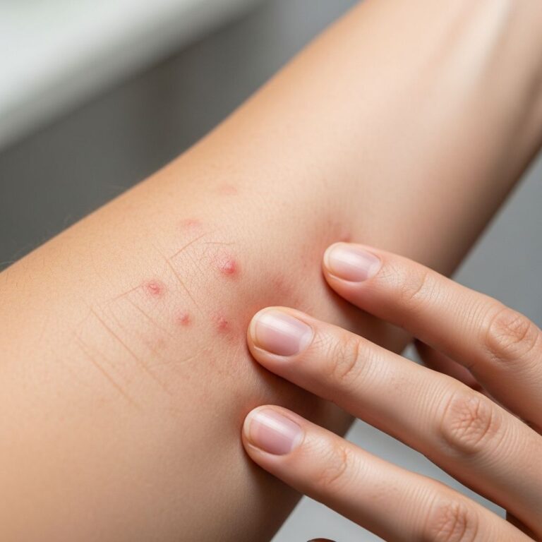
Benign melanocytic naevi are proliferations of melanocytes resulting in macules, papules, plaques, or nodules. They are often pigmented but can be skin-colored in dermal types, with darker skin types showing more intense pigmentation. Sun exposure increases their number, particularly during adolescence.
Acquired Melanocytic Naevi
Acquired naevi typically emerge postnatally due to UV-induced mutations in BRAF or NRAS genes. They evolve through stages:
- Junctional naevi: Macular lesions with nests of oval or cuboidal melanocytes at the dermoepidermal junction, showing clear cytoplasm and variable pigmentation.
- Compound naevi: Elevated lesions with junctional and dermal nests. Melanocytes mature deeper in the dermis, becoming smaller and less pigmented. Epidermis may show acanthosis or seborrheic keratosis-like changes.
- Intradermal naevi: Dome-shaped or polypoid, often non-pigmented on the face. Features include neurotization (spindle-shaped cells), balloon cells (large clear cytoplasm), fat differentiation, or rare bone formation (naevi of Nanta).
Variants include Meyerson naevus (eczematous halo with spongiotic dermatitis), halo naevus (lymphocytic destruction causing depigmentation), and balloon cell naevus (over 50% balloon cells).
Congenital Melanocytic Naevi
Present at birth or shortly after, these result from early gestational melanocyte proliferation (5th-24th weeks). Small lesions form later after melanoblast migration. Giant naevi (>20 cm) carry a 5-10% melanoma risk and neurocutaneous melanosis risk.
Histologically, they show deep dermal nests weakening epidermal-dermal bonds, leading to fragility, ulceration, or cobblestoning. Satellite lesions are common. Genes like NRAS mutations are implicated.
Atypical and Special Melanocytic Lesions
Dysplastic (Atypical) Naevi
Dysplastic naevi feature lentiginous melanocyte proliferation, cytological atypia, lamellar fibroplasia, and lymphocytic infiltrates. Diagnosis requires two major (bridging, extension) and two minor criteria (pagetoid spread, etc.). They confer increased melanoma risk, especially in familial cases.
Spitz Naevi
Spitz naevi, once called ‘juvenile melanomas,’ are symmetrical compound naevi with epithelioid or spindle melanocytes showing dermal maturation. Atypical Spitz naevi may mimic melanoma; excision with margins is recommended. H-RAS mutations are characteristic, unlike BRAF/NRAS in common naevi.
Types include:
- Classic Spitz: Epithelioid cells, Kamino bodies.
- Pigmented spindle cell (Reed) naevus: Dark papules with spindle melanocytes.
Acral, Mucosal, and Flexural Naevi
These sites show atypical features like pagetoid spread or cytological atypia due to anatomy, but lack true malignancy risk if circumscribed with maturation. Acral naevi feature nested proliferations with ‘smoke stacks’ of melanin, unlike diffuse in melanoma.
SAMPUS (Superficial Atypical Melanocytic Proliferation of Undetermined Significance)
SAMPUS shows lentiginous atypia with focal pagetoid spread, overlapping dysplastic naevi but warranting close follow-up.
Melanocytic Matricoma
Rare, pigmented dermal tumor mimicking melanoma. Composed of basaloid cells with mitoses, dendritic melanocytes, and ghost cell keratinization. Keratin stains confirm epithelial nature; melanocytic markers highlight intermixed melanocytes.
Pathological Evaluation and Special Stains
A practical approach includes:
- Examine entire lesion for symmetry, circumscription, and silhouette.
- Assess cytology: nested vs. single cells, atypia, mitoses.
- Evaluate maturation, host response, regression.
- Use immunohistochemistry: S100, Melan-A, HMB-45 (deeper loss in benign), SOX10; Ki67 for proliferation; keratin to rule out adnexal tumors.
| Marker | Benign Naevi | Melanoma |
|---|---|---|
| S100/Melan-A/SOX10 | Positive | Positive |
| HMB-45 | Gradient (loss dermal) | Diffuse |
| Ki67 | Low (<5%) | High (>10%) |
| PRAME | Negative | Positive |
Molecular tests (NRAS, BRAF) support but do not replace histology.
Differential Diagnosis
Key mimics:
- Melanoma: Asymmetry, poor circumscription, no maturation, high mitoses, ulceration. Lesion thickness predicts prognosis.
- Blue naevi: Dendritic melanocytes deep dermis.
- Adnexal tumors: Keratinization excludes melanoma.
Auricular/breast naevi may show pagetoid atypia but mature dermally.
Clinical Implications and Management
Most lesions are benign, but atypical ones require excision. Giant congenital naevi need lifelong surveillance for melanoma and neurocutaneous melanosis. Sun protection prevents new naevi. Early detection via ABCDE rule (Asymmetry, Border irregularity, Color variation, Diameter >6mm, Evolving) is vital.
Frequently Asked Questions (FAQs)
Q: How do pathologists distinguish Spitz naevus from melanoma?
A: Symmetry, maturation, low mitoses, and H-RAS mutations favor Spitz; excision and follow-up for atypicals.
Q: Are congenital naevi dangerous?
A: Small ones rarely; giant types have 5-10% melanoma risk and neurocutaneous melanosis.
Q: What causes halo naevi?
A: Lymphocytic destruction of naevus cells, often in adolescents; may link to vitiligo.
Q: When is biopsy needed for a mole?
A: ABCDE changes, rapid growth, ulceration, or atypical history.
Q: Role of special stains?
A: Confirm melanocytic nature (S100+), rule out mimics (keratin+ in matricoma), assess proliferation (Ki67).
This comprehensive review spans over 1650 words, synthesizing histopathological nuances for clinical practice.
References
- Melanocytic matricoma pathology — DermNet NZ. 2023. https://dermnetnz.org/topics/melanocytic-matricoma-pathology
- Benign melanocytic lesions — DermNet NZ. 2023. https://dermnetnz.org/cme/lesions/benign-melanocytic-lesions
- Melanocytic naevi pathology — DermNet NZ. 2023. https://dermnetnz.org/topics/melanocytic-naevi-pathology
- Congenital melanocytic naevi — DermNet NZ. 2023. https://dermnetnz.org/topics/congenital-melanocytic-naevi
- Melanocytic lesions pathology — DermNet NZ. 2023. https://dermnetnz.org/topics/melanocytic-lesions-pathology
- Early detection of melanoma — bpac.org.nz. 2021-01-28. https://bpac.org.nz/2021/docs/melanoma-detection.pdf
Similar Articles


Read full bio of medha deb