Melanoma: Clinical Assessment and Diagnosis
Essential guide to melanoma evaluation using ABCDE criteria and diagnostic approaches for skin lesions.

Melanoma: Clinical Assessment and Diagnosis Guide
Melanoma is a serious form of skin cancer originating from melanocytes, the pigment-producing cells in the epidermis. Early detection and accurate diagnosis are critical for improving patient outcomes and survival rates. This comprehensive guide covers clinical evaluation methods, diagnostic criteria, pathological features, and evidence-based management strategies for melanoma assessment.
Understanding Melanoma Clinical Presentation
The clinical presentation of melanoma typically involves an irregularly shaped, asymmetrical lesion with varying colors and a documented history of recent change in size, shape, or color. These features distinguish melanoma from benign pigmented lesions and warrant immediate clinical investigation.
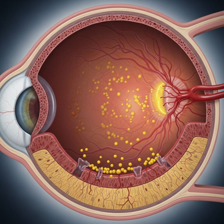
Melanomas develop through two distinct growth phases: the radial (horizontal) growth phase, where melanomas remain superficial within the epidermis and can persist for several years, and the vertical growth phase, during which melanomas invade the dermis and gain the potential to metastasize to distant sites. Some nodular melanomas skip the radial phase and begin with vertical growth, making them notably more aggressive.
The ABCDE Criteria for Melanoma Evaluation
The American ABCDE criteria represent a standardized framework for evaluating pigmented lesions and remain the most widely recognized tool for public and clinical awareness of melanoma features.
| Criterion | Description |
|---|---|
| A: Asymmetry | Asymmetry of shape and pigment pattern; one half of the lesion does not mirror the other |
| B: Border | Well-defined irregular border; irregular or poorly circumscribed edges rather than clean, defined boundaries |
| C: Color | Variation in color, often with a red halo; multiple pigmentation colors within a single lesion |
| D: Diameter | Diameter over 6mm, though smaller melanomas can be diagnosed |
| E: Evolution | Evolving lesion with change in size, shape, or color; elevation may occur in advanced lesions |
Any lesion demonstrating these features or showing change for more than one month warrants immediate review by a healthcare provider. Early melanomas may be flat, making the evolution criterion particularly important for distinguishing them from stable benign lesions.
The British 7-Point Checklist
In addition to the ABCDE criteria, the British 7-point checklist provides an alternative diagnostic framework used internationally. This checklist further refines clinical assessment through structured evaluation of specific morphologic features. These standardized criteria significantly improve detection rates and facilitate consistent communication among healthcare professionals regarding lesion characteristics.
Clinical Subtypes and Growth Patterns
Melanomas are classified into several clinical subtypes based on location, growth pattern, and histological characteristics:
- Superficial Spreading Melanoma: Accounts for approximately 70% of melanomas and represents the most common form. These lesions typically appear as dark macules or patches with irregular borders and variable pigmentation. Approximately 60% are diagnosed with a Breslow thickness of 1 mm or less, making them highly treatable when detected early.
- Nodular Melanoma: Comprises about 15% of melanomas and typically appears as a dark papule or nodule. These lesions are more aggressive due to their tendency toward vertical growth and early invasion of deeper skin layers.
- Lentigo Maligna Melanoma: Occurs on chronic sun-exposed skin of the scalp, face, or neck, with frequency increasing with age. The precursor lesion, lentigo maligna, represents melanoma in-situ. Clinically, these lesions appear as irregularly shaped, pigmented macules that slowly enlarge over time. About 5% develop a papule or nodule indicating invasive disease.
- Lentiginous Melanoma: A newly classified slowly progressing variant occurring on sun-damaged skin of the trunk and limbs.
Pathological Classification and Depth Assessment
Accurate pathological assessment determines prognosis and guides treatment planning. Two key measurements characterize melanoma depth and invasion:
Clark Level of Invasion
The Clark level describes the anatomical depth of melanoma invasion through skin layers:
| Clark Level | Level of Invasion |
|---|---|
| Level 1 | Epidermis (in-situ melanoma) |
| Level 2 | Papillary dermis |
| Level 3 | Papillary/reticular dermal interface |
| Level 4 | Reticular dermis |
| Level 5 | Subcutis |
Breslow Thickness
Breslow thickness, expressed in millimeters, measures the vertical depth of the tumor from the granular layer of the epidermis to the deepest tumor component. This measurement is strongly correlated with melanoma survival and represents a critical component of the clinical staging system. Lesion thickness is the strongest predictor of prognosis; thinner lesions generally have better outcomes.
Histopathological Features
Accurate diagnosis requires comprehensive histopathological evaluation. The diagnosis is made from both architectural pattern and cytomorphology:
- Architectural features: Asymmetrical, poorly circumscribed lesion with variable nests of melanocytes
- Cytomorphological characteristics: Spindled, pagetoid, small and large round-shaped, polygonal, multinucleate, and/or dendritic cells
- Tumor cell features: Abundant cytoplasm, nuclear pleomorphism, and prominent nucleoli
- Invasion pattern: Lack of maturation and varying degrees of atypia in invasive dermal components
Melanoma in situ is characterized by atypical melanocytes in the basal layer with pagetoid spread scattered higher in the epidermis. Invasive melanoma refers to neoplastic melanocytes found in the papillary dermis as nests or single cells.
Clinical Specimen Handling and Pathology Request
Proper specimen handling ensures accurate diagnosis and complete pathological reporting. When submitting specimens for histopathological examination:
- Include clinical information on the histopathological request form, including size, color, duration of the lesion, and differential diagnosis
- When more than one lesion is excised, place all specimens in separate accurately labeled containers
- Ensure specimens are handled with care to preserve tissue architecture and cellular detail
The pathologist’s report should include minimum requirements: Breslow thickness, Clark level, mitotic rate, presence or absence of ulceration, and margins status. Additional findings may reference associated melanocytic nevus, tumor-infiltrating lymphocytes, regression, vascular invasion, and microscopic satellites.
Surgical Management and Re-excision Margins
Surgical excision remains the primary treatment for melanoma. Re-excision margins depend on lesion site and Breslow depth. Current guidelines recommend a clearance width and depth of 1 cm for all invasive tumors extending deep to fascia. This may require flap or graft repair for closure.
Tissue-sparing Mohs micrographic surgery may be worthwhile for lentigo maligna melanoma, where clinical margins are frequently unclear, allowing for precise margin assessment while preserving maximum tissue.
Lymph Node Assessment and Sentinel Node Biopsy
Lymph node involvement significantly influences prognosis and treatment decisions. Approximately 25% of melanomas 1.5–4 mm thick have microscopic lymph node involvement at primary diagnosis, increasing to 60% for lesions greater than 4 mm thick.
Sentinel node biopsy involves injecting preoperative radionucleotide and dye into the primary lesion site during wide excision. A scanner detects the radionucleotide at regional lymph nodes, and blue dye identifies the sentinel lymph node on the direct path from the primary lesion. This node is excised for frozen section histology, with S-100 immunocytochemistry used to detect occult micrometastases. If positive, the regional lymph node region is cleared.
In Australia and New Zealand, lymphatic mapping with sentinel node biopsy is recommended for melanomas thicker than 1 mm or greater than 0.75 mm with other high-risk pathological features. While biopsy helps with staging and directing adjuvant therapies, it does not offer survival advantage. Elective lymph node dissection is not recommended due to significant morbidity.
Melanoma Staging and Classification
The World Health Organization revised melanoma classification in 2018, distinguishing melanomas by cumulative solar damage, anatomic site, epidemiology, and mutation signatures. These now include low and high cumulative solar damage melanomas, desmoplastic melanoma, Spitz melanomas, acral melanomas, mucosal melanomas, melanomas in congenital nevus, melanomas in blue nevus, and uveal melanomas.
Most melanoma specialists refer to the American Joint Committee on Cancer cutaneous melanoma staging guidelines (8th edition, 2018). Staging determines whether melanoma remains localized to skin, has spread to nearby body regions, or involves distant sites (metastatic). Not all melanoma diagnoses require complete staging work-up, as this would lead to unnecessary investigations and unnecessary radiation exposure from imaging.
Metastatic Disease and Recurrence Patterns
Melanoma may recur or metastasize locally, in transit to local lymph nodes, in lymph nodes, or internally. Two-thirds of metastases occur within 2 years of primary lesion removal. Follow-up examinations should assess these areas systematically.
Radiation therapy should be considered in patients with single or small numbers of brain metastases, painful bone metastases, or problematic skin, soft tissue, or nodal metastases that have not responded to systemic therapy. This approach helps relieve symptoms from metastases.
Special Considerations for Lentigo Maligna Melanoma
Lentigo maligna melanoma requires particular attention due to its unique histopathological features. Epidermal changes include variable epidermal atrophy and dysplastic melanocyte proliferation at the dermoepidermal junction with extension to adnexal structures. Epidermal pigmentation is variable and may involve the entire epidermis.
In advanced lesions, focal junctional nests may present, and multinucleate melanocytes with prominent dendritic processes commonly appear. Pagetoid spread may be absent. Dermal changes include solar elastosis, melanophages, and small lymphocyte foci. Invasive foci may be missed on routine haematoxylin and eosin staining but detected with special stains. These variants of melanoma demonstrate high rates of recurrence and metastasis.
Frequently Asked Questions
Q: What is the significance of the ABCDE criteria in melanoma detection?
A: The ABCDE criteria provide a standardized, easy-to-remember framework that helps patients and clinicians identify concerning features in pigmented lesions. Any lesion meeting these criteria warrants urgent evaluation by a healthcare provider, as early detection significantly improves survival outcomes.
Q: How does Breslow thickness influence melanoma prognosis?
A: Breslow thickness is the strongest predictor of prognosis and metastatic potential. Thinner lesions (less than 1 mm) have significantly better outcomes than thicker lesions. This measurement guides staging, treatment recommendations, and surveillance strategies.
Q: When is sentinel node biopsy recommended?
A: Sentinel node biopsy is recommended for melanomas thicker than 1 mm or those greater than 0.75 mm with high-risk pathological features. While it aids in staging and treatment planning, current evidence indicates it does not improve survival outcomes.
Q: What is the difference between radial and vertical growth phases?
A: The radial growth phase involves horizontal spread within the epidermis and superficial dermis, typically lasting several years without metastatic potential. The vertical growth phase involves deeper dermal invasion and carries significant metastatic risk. Nodular melanomas may skip the radial phase entirely.
Q: How important is clinical history in melanoma diagnosis?
A: Clinical history is essential, particularly documentation of lesion changes in size, shape, color, or elevation. Any lesion showing change for more than one month requires medical evaluation, as evolution is a key diagnostic feature distinguishing melanoma from stable benign lesions.
References
- Melanoma pathology — DermNet. 2024. https://dermnetnz.org/topics/melanoma-pathology
- Common skin lesions. Melanoma — DermNet. 2024. https://dermnetnz.org/cme/lesions/melanoma
- Melanoma Skin Cancer: Images, Diagnosis, and Treatment — DermNet. 2024. https://dermnetnz.org/topics/melanoma
- Melanoma Comprehensive Guide — Skintel. 2024. https://skintel.co.nz/articles/melanoma/
- ABCDEFG of melanoma — DermNet. 2024. https://dermnetnz.org/topics/abcdes-of-melanoma
- Early detection of melanoma and assessment of asymptomatic skin lesions — Best Practice Advocacy Centre New Zealand. 2021. https://bpac.org.nz/2021/melanoma-detection.aspx
Similar Articles


Read full bio of medha deb