Melanoma Of The Head And Neck: Comprehensive Guide For 2025
Understanding diagnosis, treatment, and management of head and neck melanoma.

Understanding Melanoma of the Head and Neck
Melanoma is a serious form of skin cancer that develops from melanocytes, the cells responsible for producing pigment in the skin. When melanoma occurs in the head and neck region, it presents unique clinical challenges due to the complex anatomy, cosmetic concerns, and proximity to critical structures. Head and neck melanoma accounts for a significant proportion of all melanoma cases and requires specialized multidisciplinary management to achieve optimal outcomes.
The head and neck region encompasses the face, scalp, ears, neck, and surrounding tissues. Melanomas in this area tend to have different biological behaviors compared to melanomas on other body sites, including variations in lymphatic drainage patterns and metastatic potential. Understanding the specific characteristics of head and neck melanoma is essential for patients, caregivers, and healthcare providers involved in treatment planning and follow-up care.
Risk Factors and Prevention
Several risk factors increase the likelihood of developing melanoma in the head and neck region. Fair skin, light eyes, and a tendency to burn easily are significant risk factors. Individuals with a family history of melanoma or a personal history of previous skin cancers face elevated risk.
Cumulative sun exposure is one of the most important preventable risk factors. Unlike some other melanomas that are associated with intermittent, intense sun exposure and sunburns, head and neck melanomas are frequently linked to chronic, cumulative UV radiation exposure. Outdoor workers, individuals living in sunny climates, and those with recreational sun exposure patterns are at higher risk.
Additional risk factors include:
- Presence of atypical moles or dysplastic nevi
- Immunosuppression from transplant medications or chronic lymphoproliferative disorders
- Advanced age
- Male gender, particularly for scalp and neck locations
- Genetic predisposition and hereditary melanoma syndromes
Prevention strategies focus on limiting UV exposure through sun protective clothing, broad-spectrum sunscreen with SPF 30 or higher, seeking shade during peak sun hours, and avoiding indoor tanning. Regular self-examinations and professional skin surveillance for high-risk individuals are crucial for early detection.
Clinical Presentation and Diagnosis
Recognizing the early signs of melanoma is critical for achieving better treatment outcomes. The ABCDE criteria serve as a helpful guideline for identifying potentially concerning lesions:
- Asymmetry: One half of the mole does not match the other half
- Border: The edges are irregular, ragged, notched, or blurred
- Color: The lesion contains multiple colors or uneven distribution of pigment
- Diameter: The lesion is larger than 6 millimeters, approximately the size of a pencil eraser
- Evolution: The mole is changing in size, shape, color, or symptoms
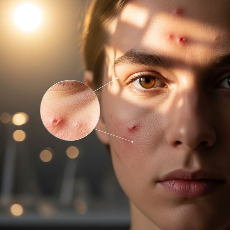
Melanomas can present in various forms. Nodular melanomas appear as raised, firm bumps and may be amelanotic, lacking significant pigmentation. Lentigo maligna melanomas typically appear as flat, irregular patches with variable pigmentation and are most common on sun-exposed areas of the face in older individuals. Superficial spreading melanomas are more common and may present with irregular borders and color variation.
Symptoms associated with melanoma include itching, bleeding, or oozing from a pigmented lesion. However, many melanomas may not cause any symptoms, emphasizing the importance of visual examination.
The diagnostic process begins with a thorough clinical history and physical examination. Dermatoscopy, a specialized imaging technique using a magnified lens with polarized light, enhances visualization of pigment patterns and structures not visible to the naked eye. Total body photography provides baseline documentation for future comparison and helps identify changing lesions.
Definitive diagnosis requires histopathological examination through biopsy. A full-thickness excisional biopsy is preferred when feasible, as it allows complete histological assessment and determines important prognostic factors. Punch or shave biopsies may be performed for initial evaluation, though excisional biopsy remains the gold standard.
Staging and Pathological Assessment
Accurate staging is essential for determining prognosis and guiding treatment decisions. The TNM (Tumor, Node, Metastasis) staging system classifies melanoma based on:
- Tumor characteristics: Thickness (measured in millimeters), ulceration, mitotic rate, and level of invasion
- Regional lymph node involvement: Number, location, and size of involved nodes
- Distant metastasis: Presence of cancer in distant organs or non-regional lymph nodes
Breslow thickness, the measurement from the granular layer to the deepest point of tumor invasion, is the most important prognostic factor for primary cutaneous melanoma. Thinner tumors (<1 mm) have significantly better prognosis, while thicker tumors (>4 mm) carry higher risk of recurrence and metastasis. Ulceration of the primary tumor indicates poor prognosis and upstages the lesion.
Molecular testing for BRAF, NRAS, and KIT mutations, as well as PD-L1 expression, provides additional prognostic and predictive information that influences treatment selection, particularly for advanced-stage disease.
Surgical Treatment
Surgery remains the primary treatment modality for localized melanoma of the head and neck. The primary goals are complete tumor removal with adequate margins while preserving function and optimizing cosmetic outcomes.
Primary Tumor Excision
Wide local excision with appropriate surgical margins is the standard treatment for melanoma. Recommended margins are based on Breslow thickness:
- In situ melanoma: 5-10 mm margins
- Melanoma 1 mm or less in thickness: 10 mm margins
- Melanoma 1-2 mm in thickness: 10-20 mm margins
- Melanoma greater than 2 mm in thickness: 20 mm margins
In the head and neck region, achieving these margins while maintaining function and appearance can be challenging. Surgeons must balance oncologic principles with cosmetic and functional considerations. Reconstruction may be necessary following excision, utilizing techniques such as skin grafts, local flaps, or regional flaps depending on the defect size and location.
Sentinel Lymph Node Biopsy
Sentinel lymph node biopsy (SLNB) is recommended for melanomas with intermediate thickness (1-4 mm) and for certain high-risk thin melanomas. This procedure involves injecting radioactive tracer and blue dye near the primary tumor to identify the first lymph node(s) to which cancer is likely to spread. The sentinel node is then surgically removed and examined for the presence of cancer cells.
SLNB provides accurate staging information and can identify occult lymph node metastasis not apparent on clinical examination. Patients with positive sentinel nodes are candidates for completion neck dissection and adjuvant systemic therapy. The head and neck region may have complex lymphatic drainage patterns, with multiple sentinel nodes in different nodal basins, requiring careful surgical planning.
Neck Dissection
Therapeutic neck dissection is performed when regional lymph node metastasis is confirmed by sentinel node biopsy or clinical examination. The type of dissection depends on the location and extent of nodal disease. Radical neck dissection removes all lymph nodes from levels I through V along with the sternocleidomastoid muscle, internal jugular vein, and spinal accessory nerve. Modified radical neck dissection preserves one or more of these non-lymphoid structures while still removing the lymph nodes. Selective neck dissection removes specific nodal levels based on the primary tumor location and lymphatic drainage patterns.
Adjuvant and Systemic Therapy
For advanced-stage melanoma or tumors with high-risk features, systemic therapy may be recommended in addition to surgery.
Immunotherapy
Immunotherapy, particularly checkpoint inhibitors targeting PD-1 and CTLA-4, has revolutionized melanoma treatment. Agents such as nivolumab, pembrolizumab, and combination regimens of nivolumab plus ipilimumab are employed for Stage III and Stage IV melanoma. These agents activate the immune system to recognize and attack melanoma cells. Immunotherapy has significantly improved survival rates for patients with advanced disease and is increasingly used as adjuvant therapy following surgery in high-risk patients.
Targeted Therapy
For melanomas with BRAF V600E or V600K mutations, BRAF inhibitors combined with MEK inhibitors provide alternative treatment options. These targeted therapies work by blocking specific mutations driving melanoma growth. Patients with BRAF-mutated metastatic melanoma may receive combinations such as dabrafenib plus trametinib or vemurafenib plus cobimetinib.
Radiation Therapy
Adjuvant radiation therapy is considered for high-risk features including thick primary tumors, close or positive surgical margins, extranodal extension from involved nodes, or extensive nodal involvement. Radiation may also be used for palliative treatment of brain metastases or other symptomatic sites of disease.
Follow-Up and Surveillance
Patients with a history of melanoma require lifelong surveillance due to the risk of recurrence and second primary melanomas. Follow-up intervals depend on stage at diagnosis and other risk factors. High-risk patients typically undergo clinical examination every 3-6 months, with imaging studies such as CT or PET scans as clinically indicated.
Regular self-examination using the ABCDE criteria is essential. Total body photography helps identify new or changing lesions. Dermatologic surveillance allows early detection of recurrent or second primary melanomas, which may be more readily curable if identified early.
Prognosis and Survival Outcomes
Prognosis for melanoma of the head and neck depends on multiple factors including Breslow thickness, ulceration, mitotic rate, stage at diagnosis, and molecular characteristics. Five-year survival rates range from over 90% for thin, early-stage melanomas to significantly lower rates for Stage IV disease with distant metastases.
Head and neck melanomas may have different prognostic implications compared to melanomas on other body sites. Certain anatomical locations, such as scalp melanomas, historically carry worse prognosis. However, advances in treatment modalities have improved outcomes across all stages.
Special Considerations: Mucosal Melanoma
Mucosal melanomas of the head and neck, occurring in the nasal cavity, paranasal sinuses, or oral cavity, represent a particularly aggressive subset. These tumors typically present at advanced stages and have poor prognosis. Surgical excision remains the primary treatment modality, though anatomical complexities can make complete excision challenging. Immunotherapy is increasingly employed for advanced mucosal melanomas, showing promise in clinical practice.
Frequently Asked Questions
Q: What are the early warning signs of head and neck melanoma?
A: Early warning signs include changes in existing moles, new pigmented lesions with irregular borders, color variation, or lesions larger than 6 mm. Any mole that itches, bleeds, or changes in appearance warrants medical evaluation.
Q: How is melanoma of the head and neck diagnosed?
A: Diagnosis requires clinical examination, dermatoscopy, and histopathological evaluation through biopsy. Additional imaging studies and molecular testing may be performed to stage the disease and guide treatment decisions.
Q: What is sentinel lymph node biopsy?
A: Sentinel lymph node biopsy is a surgical procedure that identifies and removes the first lymph node(s) to which cancer is most likely to spread. This provides important staging information and helps determine if additional treatment is needed.
Q: What are the treatment options for melanoma of the head and neck?
A: Treatment options include wide local excision with appropriate margins, sentinel lymph node biopsy, neck dissection if lymph nodes are involved, and adjuvant therapies including immunotherapy, targeted therapy, and radiation therapy. Treatment decisions are individualized based on stage, risk factors, and patient preferences.
Q: How often should I have follow-up visits after melanoma treatment?
A: Follow-up intervals depend on your stage and risk factors. High-risk patients typically have visits every 3-6 months initially, with intervals that may be extended over time. Lifelong surveillance is recommended due to the risk of recurrence and second primary melanomas.
Q: Can melanoma of the head and neck be prevented?
A: While not all melanomas can be prevented, risk can be reduced by limiting UV exposure, wearing sun protective clothing and sunscreen, avoiding indoor tanning, and having regular skin surveillance if you have risk factors.
References
- Management Controversies in Head and Neck Melanoma — National Center for Biotechnology Information (NCBI). 2016. https://pubmed.ncbi.nlm.nih.gov/27606893/
- Current management of mucosal melanoma of the head and neck — Journal of Surgical Oncology, Johns Hopkins University. 2003-06-01. https://pure.johnshopkins.edu/en/publications/current-management-of-mucosal-melanoma-of-the-head-and-neck
- Overview of the Hopkins Melanoma/Skin Cancer Program — Johns Hopkins Medicine. 2024. https://www.hopkinsmedicine.org/
- The Current State of Metastatic Melanoma — ECOG-ACRIN Cancer Research Group, Sidney Kimmel Comprehensive Cancer Center at Johns Hopkins Medicine. 2024. https://ecog-acrin.org/the-current-state-of-metastatic-melanoma/
- National Comprehensive Cancer Network (NCCN) Guidelines for Melanoma — NCCN. 2025. https://www.nccn.org/
Similar Articles


Read full bio of medha deb