Melanoma Pathology: Complete Guide To Diagnosis & Staging
Comprehensive guide to the histopathological features, subtypes, and prognostic factors of melanoma skin cancer.

Melanoma is a malignant neoplasm originating from melanocytes, the pigment-producing cells in the skin. Pathological examination is essential for definitive diagnosis, subtyping, and determining prognosis through assessment of growth phases, invasion depth, and cellular features.
Who is at Risk?
Individuals with fair skin, multiple nevi, history of sunburns, and genetic predispositions such as mutations in CDKN2A are at higher risk for melanoma. Chronic sun exposure and UV radiation play critical roles in pathogenesis, leading to DNA damage and activation of pathways like MAPK, with common mutations in BRAF (45%) and NRAS (18%).
Clinical Features
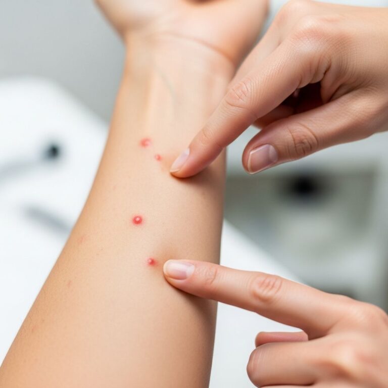
Melanomas often present as asymmetrical lesions with irregular borders, varied colours, and diameters greater than 6 mm (ABCDE rule). Subtypes vary: superficial spreading melanoma appears as irregular macules or patches, while nodular melanoma presents as rapidly growing dark nodules. Dermoscopy may reveal atypical pigment networks, blue-white veils, and irregular globules.
What are the Growth Phases of Melanoma?
Most melanomas exhibit an initial
radial growth phase
confined to the epidermis and sometimes papillary dermis, followed by avertical growth phase
with dermal invasion. Nodular melanomas often bypass or rapidly overrun the radial phase, presenting directly with vertical growth.- Radial growth phase: Horizontal spread within the epidermis, low metastatic potential.
- Vertical growth phase: Invasion into dermis, increased risk of metastasis.
How is the Diagnosis Made?
Diagnosis requires histopathological examination of a biopsy. Suspicious lesions should be excised with clear margins where possible. Pathologists assess architectural disorder, cytological atypia, and invasion depth.
What are the Histological Features?
Melanomas are asymmetrical, poorly circumscribed lesions with marked atypia. Key features include:
- Consumption of the epidermis (thinning over tumour nests).
- Pagetoid spread of atypical melanocytes upward into the epidermis.
- Nests of melanocytes of variable size/shape, often confluent, lacking maturation with depth.
- Melanocytes in lymphovascular spaces.
- Deep and atypical mitoses, increased apoptosis.
- Ulceration, a poor prognostic factor.
Tumour cells show abundant cytoplasm, nuclear pleomorphism, and prominent nucleoli. Mitotic figures are common.
Clark Level and Breslow Thickness
Prognostic staging uses Clark level (anatomical invasion level) and Breslow thickness (tumour depth in mm from granular layer to deepest point).
| Clark Level | Level of Invasion |
|---|---|
| 1 | Epidermis (in-situ melanoma) |
| 2 | Papillary dermis |
| 3 | Papillary/reticular dermal interface |
| 4 | Reticular dermis |
| 5 | Subcutis |
Breslow thickness strongly correlates with survival and is integral to AJCC staging. Thinner lesions (≤1 mm) have better outcomes.
Superficial Spreading Melanoma
The most common subtype (70%), often on intermittently sun-exposed skin. Features include:
- Proliferation of melanocytes along dermoepidermal junction, extending over rete ridge tips.
- Variable nests, pagetoid spread, epidermal consumption.
- Invasive dermal component with lack of maturation and atypia.
Melan-A immunostaining highlights atypical melanocytes.
Nodular Melanoma
Accounts for 15%, presents as rapidly enlarging nodule. Histology shows a symmetrical dermal mass of dysplastic cells with upward epidermal invasion, minimal radial phase.
Lentigo Maligna Melanoma
Occurs on chronically sun-exposed areas (face, scalp) in older individuals. Precursor is lentigo maligna (melanoma in-situ). Features:
- Irregular pigmented macule with slow enlargement.
- Epidermal atrophy, dysplastic melanocytes at junction extending to adnexa.
- Variable pigmentation, occasional junctional nests, multinucleate melanocytes.
- Dermal solar elastosis, melanophages; invasive foci detected by special stains like Melan-A.
Invasive component shows spindle or epithelioid cells.
Lentiginous Melanoma
Slowly progressing on sun-damaged trunk/limbs. Histology: lentiginous hyperplasia, focal junctional nests, cytological atypia, pagetoid spread.
Desmoplastic Melanoma
Rare, spindle-shaped tumour cells in dermis/subcutis amid collagen bundles. Often deeply infiltrative; special stains needed for depth assessment. May mimic dermatofibroma; lymphocytes/plasma cells aid diagnosis.
What is the Role of Immunohistochemistry?
Immunostains confirm melanocytic origin and aid in difficult cases:
- S100: Sensitive for neural crest origin, positive in melanoma/desmoplasia.
- HMB-45 (gp100): Highlights radial growth phase, diminishes with depth.
- Melan-A (MART-1): Highly sensitive/specific, delineates lesions and invasion.
- Tyrosinase, SOX10: Additional markers for confirmation.
Ki67 assesses proliferation; useful in spitzoid lesions.
Other Rare Melanomas
WHO classification includes acral, mucosal, uveal melanomas, and those arising in congenital/blue nevi, distinguished by site, CSD, and mutations.
Melanoma Staging
Uses AJCC 8th edition TNM system based on Breslow thickness, ulceration, mitoses, nodal/distant metastasis. Staging guides treatment; not all cases need full workup to avoid unnecessary scans.
Frequently Asked Questions (FAQs)
What is the strongest predictor of melanoma prognosis?
Breslow thickness; thinner lesions (≤1 mm) are highly treatable with excellent outcomes.
How does nodular melanoma differ histologically?
It shows a dominant vertical growth phase with dermal tumour mass and minimal radial phase.
What does consumption of the epidermis mean?
Thinning of epidermis over atypical melanocyte nests, a hallmark of melanoma.
Why use special stains like Melan-A?
To detect subtle invasion, delineate margins, especially in lentigo maligna or desmoplastic types.
Is Clark level still used?
Less than Breslow, but provides anatomical context in pathology reports.
References
- Melanoma Pathology — DermNet NZ. 2023. https://dermnetnz.org/topics/melanoma-pathology
- Melanoma Comprehensive Guide — Skintel. 2023. https://skintel.co.nz/articles/melanoma/
- Melanoma Skin Cancer — DermNet NZ. 2023. https://dermnetnz.org/topics/melanoma
- Malignant Melanoma of the Skin — Geeky Medics. 2023-10-01. https://geekymedics.com/malignant-melanoma-of-the-skin/
- New Zealand Melanoma Clinical Guidelines — Melanoma Network of New Zealand. 2023. https://melnet.org.nz/assets/DRAFT-Melanoma-Clinical-Guidelines-for-consultation.pdf
Similar Articles


Read full bio of medha deb