Merkel Cell Carcinoma: Causes, Symptoms, and Treatment
Understanding MCC: A comprehensive guide to diagnosis, treatment, and prognosis.

Merkel Cell Carcinoma
Merkel cell carcinoma (MCC) is a rare and highly aggressive cutaneous neuroendocrine neoplasm that arises from uncontrolled growth of cells in the skin. This malignancy primarily affects elderly individuals and those with compromised immune function, and it carries a significantly higher mortality rate compared to melanoma. Despite its rarity, MCC presents substantial clinical challenges due to its rapid growth potential, high propensity for metastasis, and the limited availability of established treatment guidelines.
Definition and Overview
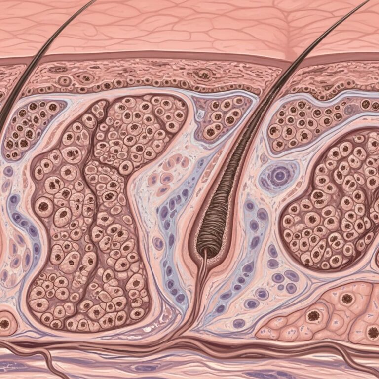
Merkel cell carcinoma is a rare cutaneous neuroendocrine malignancy that originates from Merkel cells, specialized sensory cells located in the basal layer of the epidermis involved in touch sensation. MCC is characterized by its aggressive biological behavior, with a high risk of regional and distant metastases. The disease typically manifests as an asymptomatic, rapidly growing nodule on sun-exposed skin areas, frequently leading to initial misdiagnosis as a benign lesion. The case fatality rate for MCC exceeds that of melanoma, underscoring the importance of early recognition and appropriate management.
Epidemiology and Risk Factors
Merkel cell carcinoma predominantly affects white adults with advancing age, with the majority of cases occurring in individuals over 50 years old. The disease shows a marked male predominance and is rare before the fifth decade of life. Several well-established risk factors contribute to MCC development:
- Cumulative ultraviolet (UV) radiation exposure from sun exposure is a major contributor, particularly in individuals with light skin types
- Immunosuppression significantly elevates risk, including patients with HIV/AIDS, those receiving immunosuppressive medications for organ transplantation, and individuals with chronic lymphoproliferative disorders
- Merkel cell polyomavirus (MCPyV) infection with oncogenic transformation represents a distinct etiologic pathway, particularly in immunocompromised populations
- Advanced age correlates strongly with increased incidence
- Male sex confers approximately twofold increased risk compared to females
- White skin type shows highest susceptibility
A useful clinical mnemonic, AEIOU, summarizes typical characteristics: Asymptomatic, Expanding rapidly, Immune suppressed, Older than 50 years, and UV-exposed skin. Research demonstrates that 89% of patients meet three or more criteria, while 52% fulfill four or more elements of this mnemonic.
Clinical Presentation and Diagnosis
History and Examination
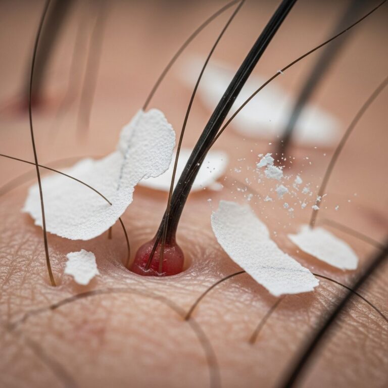
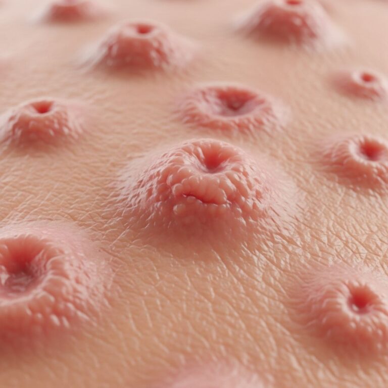
The primary MCC tumor typically presents as an asymptomatic, rapidly growing, firm, nontender papule or subcutaneous nodule on sun-exposed skin. The lesion characteristically appears pink, violaceous, or skin-colored and is often located on the face, neck, ears, or extremities. Key diagnostic features include:
- Rapidly expanding lesion over weeks to months
- Firm consistency without tenderness
- Red, pink-violaceous, or skin-colored appearance
- Location on sun-exposed areas
- Dermal or subcutaneous depth
- Possible ulceration or bleeding in advanced lesions
- Satellite papules or nodules surrounding the primary tumor
- Enlarged regional lymph nodes suggesting nodal involvement
Diagnostic Testing
Skin biopsy with histopathologic assessment remains the gold standard for MCC diagnosis. A tissue sample obtained through punch or excisional biopsy is examined microscopically to confirm the presence of characteristic MCC cells. Immunohistochemical staining for neuroendocrine markers such as synaptophysin, chromogranin, and cytokeratin 20 supports the diagnosis and helps differentiate MCC from mimics including lymphoma, metastatic small cell carcinoma, and other neuroendocrine malignancies.
Dermoscopy may reveal helpful features but should not delay tissue diagnosis. Additional diagnostic modalities include:
- Lymph node ultrasound to assess regional nodal involvement
- Whole-body positron emission tomography (PET) scan for metastatic staging
- Contrast-enhanced CT imaging of the chest, abdomen, and pelvis to evaluate for distant disease
- Brain MRI in selected cases with concerning clinical features
- Sentinel lymph node biopsy (SLNB) to determine pathologic nodal status
- Merkel cell polyomavirus serology to assess MCPyV exposure
- Circulating tumor DNA (ctDNA) analysis as an emerging prognostic marker
Staging and Histologic Subtypes
Merkel cell carcinoma staging utilizes the American Joint Committee on Cancer system, incorporating primary tumor size, presence of regional lymph node metastases, and distant metastatic disease:
| Stage | Description | Five-Year Survival |
|---|---|---|
| Stage I | Localized tumor (≤2 cm) with no lymph node involvement | 50-80% |
| Stage II | Larger primary tumor (>2 cm) with no lymph node involvement | 50-80% |
| Stage IIIA | Tumor with clinically detected regional lymph node metastases | 30-60% |
| Stage IIIB | Tumor with pathologically confirmed lymph node metastases | 30-60% |
| Stage IV | Distant metastatic disease to organs such as lungs or liver | <20% |
Histologically, MCC demonstrates three distinct subtypes: trabecular (classic large-cell pattern with high-density granules), intermediate (solid pattern, most common), and small cell (diffuse pattern with few high-density granules). Tumor size represents the most significant prognostic parameter, with larger tumors correlating with increased metastatic risk. However, MCC tumors of any size carry substantial risk for occult metastasis, supporting the use of sentinel lymph node biopsy for all cases.
Treatment Approaches
Localized Disease (Stages I-II)
Management of localized MCC typically involves surgical wide local excision with adequate margins of healthy tissue surrounding the tumor. This approach aims to minimize recurrence risk while preserving function and cosmesis. Mohs micrographic surgery may be performed in cosmetically or functionally sensitive areas such as the face or eyelid, allowing precise removal of cancerous tissue while maximizing healthy tissue preservation through microscopic layer-by-layer examination during surgery.
Sentinel lymph node biopsy should be performed during primary tumor surgery to determine pathologic nodal status, as this information significantly impacts staging and treatment decisions. Following surgery, adjuvant radiation therapy to the primary site and regional nodal basin is frequently recommended due to MCC’s apparent radiosensitivity and the high incidence of local and regional recurrences after surgery alone. Radiation is particularly important for patients with larger tumors, close or positive excision margins, locally unresectable tumors, or positive regional lymph nodes.
Regional Nodal Disease (Stage III)
Treatment of stage III MCC with regional lymph node involvement requires a multidisciplinary approach. Options include radiation therapy to the nodal basin, lymph node dissection, or a combination of both modalities. Neoadjuvant immunotherapy with immune checkpoint inhibitors may be considered prior to surgery or radiation to reduce tumor burden and improve outcomes. The specific approach should be individualized based on patient factors, nodal disease extent, and patient preferences regarding treatment burden and morbidity.
Distant Metastatic Disease (Stage IV)
For patients with stage IV metastatic MCC, enrollment in a clinical trial represents the preferred treatment option according to both US and European guidelines. When clinical trials are unavailable, treatment may involve any combination of the following: immunotherapy using immune checkpoint inhibitors (such as pembrolizumab or avelumab), radiation therapy to sites of metastatic disease, surgical resection of limited metastatic lesions, or conventional chemotherapy. The aggressive nature of stage IV disease necessitates prompt treatment initiation and close clinical monitoring.
Prognosis and Surveillance
Merkel cell carcinoma carries a considerably worse prognosis than cutaneous melanoma, with survival rates dependent on stage at diagnosis. Early-stage localized disease demonstrates five-year survival rates of 50-80%, while regional nodal involvement reduces survival to 30-60%. Distant metastatic disease carries the poorest prognosis, with five-year survival rates typically below 20%.
MCC recurs in up to 50% of patients, necessitating rigorous ongoing surveillance. Patients require regular clinical examination of the primary tumor site and regional lymph node basins, with imaging studies as clinically indicated. Regular dermatologic follow-up appointments are essential for detecting recurrent or new primary tumors at the earliest opportunity when treatment may be most effective.
Frequently Asked Questions
Q: How is Merkel cell carcinoma different from melanoma?
A: While both are skin cancers, MCC is a neuroendocrine malignancy with a higher mortality rate than melanoma. MCC typically presents as a rapidly growing nodule that is often asymptomatic and frequently misdiagnosed initially. MCC carries greater risk of metastasis and demonstrates more aggressive biological behavior than most melanomas.
Q: What is the role of Merkel cell polyomavirus in MCC development?
A: MCPyV represents a distinct etiologic pathway for MCC, particularly in immunocompromised individuals. The virus undergoes oncogenic transformation, contributing to tumor development. This represents one of two major recognized etiologies alongside UV radiation exposure, with some tumors arising from both mechanisms.
Q: Should all patients with MCC undergo sentinel lymph node biopsy?
A: Yes, sentinel lymph node biopsy should be performed for all MCC patients, as MCC tumors of any size carry substantial risk for occult lymph node metastasis. The pathologic nodal status significantly impacts staging, prognosis, and treatment decisions.
Q: Is immunotherapy effective for advanced MCC?
A: Immunotherapy with immune checkpoint inhibitors has emerged as an important treatment option for advanced MCC, particularly in the metastatic setting. Clinical trials demonstrating efficacy have led to guideline recommendations for immunotherapy as a primary treatment approach for stage IV disease.
Q: What is the importance of early detection in MCC?
A: Early detection dramatically improves outcomes, with localized disease demonstrating substantially better survival rates (50-80% five-year survival) compared to advanced stages. Awareness of MCC’s characteristic rapid growth on sun-exposed skin in elderly or immunocompromised individuals enables prompt diagnosis and treatment initiation.
References
- Merkel cell carcinoma – Symptoms, diagnosis and treatment — BMJ Best Practice. 2025. https://bestpractice.bmj.com/topics/en-us/3000356
- Merkel Cell Carcinoma Treatment (PDQ®) — National Cancer Institute. 2025. https://www.cancer.gov/types/skin/hp/merkel-cell-treatment-pdq
- Merkel cell carcinoma – Diagnosis and treatment — Mayo Clinic. 2024. https://www.mayoclinic.org/diseases-conditions/merkel-cell-carcinoma/diagnosis-treatment/drc-20351036
- Merkel Cell Carcinoma of the Skin — StatPearls, National Center for Biotechnology Information. 2024. https://www.ncbi.nlm.nih.gov/books/NBK482329/
- NCCN Guidelines® for Merkel cell carcinoma — National Comprehensive Cancer Network. 2025. https://merkelcell.org/wp-content/uploads/2025/06/NCCN_MCC_2025Guidelines.pdf
- Merkel Cell Carcinoma Guide for Patients & Providers — Merkel Cell Carcinoma Research Foundation. 2025. https://merkelcell.org/wp-content/uploads/2025/08/2025-MCC-Handout.pdf
Similar Articles


Read full bio of Sneha Tete