Merkel Cell Carcinoma Pathology Guide: Key Diagnosis Insights
Comprehensive pathology guide to Merkel cell carcinoma: clinical features, histopathology, diagnosis, and management insights.

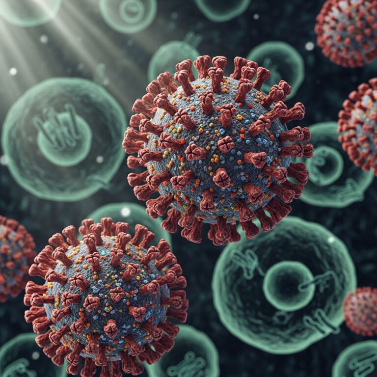
Merkel cell carcinoma (MCC) is a rare, aggressive neuroendocrine carcinoma arising primarily from the skin, characterized by rapid growth and high metastatic potential. It predominantly affects sun-exposed areas in elderly individuals and is associated with Merkel cell polyomavirus (MCV) integration or UV-induced mutations.
Introduction
Merkel cell carcinoma represents a highly malignant skin tumor originating from neuroendocrine cells, with an incidence linked to immunosuppression, advanced age, and UV exposure. The tumor’s pathology reveals distinct subtypes based on viral etiology or genetic damage, influencing morphology, behavior, and prognosis. Approximately 80% of cases harbor MCV, showing better outcomes compared to virus-negative tumors driven by TP53 mutations and RB1 alterations.
Clinical Features
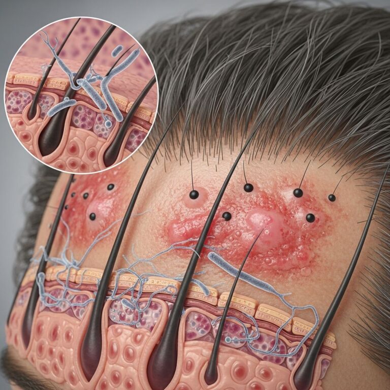
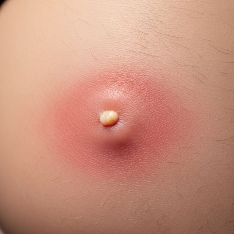
MCC typically presents as a rapidly expanding, painless, firm nodule on sun-damaged skin, most commonly on the head, neck, or extremities. Lesions are often flesh-colored, erythematous, or violaceous, measuring 0.5–5 cm, with possible ulceration, scale, or telangiectasia. The AEIOU criteria aid diagnosis: Asymptomatic, Expanding rapidly, Immunosuppressed, Older than 50, UV-exposed site, met by 89% of patients.
- Dome-shaped nodule: Firm, nontender, 1–2 cm at diagnosis.
- Color variation: Reddish-purple hue common.
- Satellite lesions: Frequent in advanced cases.
- Lymph node involvement: Early regional metastasis in 10–30%.
Prognosis correlates with stage: localized disease has 70–80% 5-year survival, dropping to 52% with nodal involvement.
Epidemiology
MCC incidence has risen from 0.15 to 0.44 per 100,000 in the US (1986–2001), with highest rates in fair-skinned elderly males. Risk factors include UV exposure, immunosuppression (HIV, transplant), and MCV infection. Head/neck accounts for 36–50% of cases, limbs 40%.
| Clinical Features | Dermoscopic Features |
|---|---|
| Nonspecific dome-shaped firm nodule or tumor Flesh-colored to erythematous to violaceous +/− overlying scale and telangiectasia | Polymorphic vascular pattern Milky-white structureless areas |
Histopathology
MCC arises in the dermis, extending to subcutaneous fat, sparing or ulcerating the epidermis. Tumor cells form large subepidermal nests or trabeculae with scant cytoplasm, round/oval nuclei, vesicular chromatin, prominent nucleoli, high mitotic rate, and apoptosis. Three subtypes exist: intermediate (most common), small cell, trabecular—often admixed, without prognostic difference. Lymphovascular invasion is nearly universal.
- Tumor architecture: Sheet-like, trabecular, or nested patterns.
- Cytology: Small blue round cells, molding, crush artifact.
- Stroma: Desmoplastic in some cases.
- Variants: Rare keratinization or ductal differentiation.
Electron microscopy shows dense-core neurosecretory granules (80–200 nm) and paranuclear keratin aggregates, confirming neuroendocrine origin.
Cytogenetics and Molecular Pathology
Two pathogenic subsets: MCV-positive (80%, better prognosis) with viral T-antigen expression sequestering RB1; MCV-negative (UV-driven) with TP53 mutations, RB1 deletion/hypermethylation. Chromosomal losses at 1p36, 3p, 10q, 13q; gains at 1q, 3q, 5p, 6p, 8q, 18q. MCV integration is clonal, distinguishing primary MCC from metastases.
Immunohistochemistry
IHC is pivotal for diagnosis, showing perinuclear dot-like CK20 positivity (95%), CK8/18 (CAM5.2), neurofilament (paranuclear globules), and neuroendocrine markers: synaptophysin, chromogranin A, NSE. Negative for S100, HMB45, TTF1 (vs. lung small cell), SOX10. PD-L1 expression common in MCV+ cases, guiding immunotherapy.
| Positive Markers | Negative Markers |
|---|---|
| CK20 (dot-like) Synaptophysin Chromogranin NSE Neurofilament | S100 TTF1 SOX10 TTF1 (lung SCC) |
Differential Diagnosis
MCC mimics other small round blue cell tumors. Distinction relies on IHC and clinical context.
| Differential | Distinguishing Features | IHC |
|---|---|---|
| Basal cell carcinoma (BCC) | Peripheral palisading, retraction artifact, mucin | BerEP4+, CK20-, MCV- |
| Small cell lung carcinoma (SCLC) | Pulmonary history, TTF1+ | TTF1+, CK20- |
| Melanoma | Pigmentation, junctional activity | S100+, SOX10+, MELK- |
| Lymphoma | Clonal lymphoid markers | CD45+, CD20/CD3+ |
| Sebaceous carcinoma | Multivacuated cells, lipid | Embryonal antigen+ |
Staging and Prognosis
Staging uses AJCC 8th edition: Stage 0 (in situ), I (≤2cm, node-), II (>2cm or local invasion), III (nodal), IV (distant). Prognosis: 5-year survival 62% overall; 97% node-negative vs. 52% node-positive. MCV+ better than MCV-. Recurrence in 40%, often within 12 months; metastases to nodes (first), then skin, lung, bone, brain.
- Favorable: Head/neck primary, smaller size, MCV+.
- Poor: Trunk/extremity, lymphovascular invasion, node+.
Treatment Overview
Surgery (wide excision, 1–2cm margins) with SLNB for staging. Adjuvant RT for high-risk; immunotherapy (anti-PD-1/PD-L1) for advanced/MCV+. Chemotherapy less effective.
Frequently Asked Questions (FAQs)
What is Merkel cell carcinoma?
A rare, aggressive skin cancer from neuroendocrine cells, often virus-associated.
Who is at risk for MCC?
Elderly, fair-skinned, immunosuppressed individuals with sun exposure.
How is MCC diagnosed pathologically?
By histopathology showing small blue cells and IHC (CK20 dot-like, synaptophysin+).
What is the prognosis?
Variable; 5-year survival 50–70%, worse with metastases.
Is MCC curable?
Yes, if localized and treated early with surgery and radiation.
References
- Merkel Cell Carcinoma — Skelton HG et al. PMC. 2017-05-01. https://pmc.ncbi.nlm.nih.gov/articles/PMC5443625/
- Merkel cell carcinoma: A review — Walsh NM. Wiley Online Library. 2020-01-15. https://onlinelibrary.wiley.com/doi/10.1111/cup.13910
- Merkel Cell Carcinoma — UCSF Surgical Oncology. 2025-01-01. https://surgicaloncology.ucsf.edu/condition/merkel-cell-carcinoma
- Merkel Cell Carcinoma — EyeWiki. 2024-06-01. https://eyewiki.org/Merkel_Cell_Carcinoma
- Merkel Cell Carcinoma Review — Goessling W et al. Merkel Cell Carcinoma. 2001-10-01. https://merkelcell.org/wp-content/uploads/2016/10/MCCReviewGoessling.pdf
- Merkel Cell Carcinoma of the Skin — NCBI Bookshelf. 2023-07-17. https://www.ncbi.nlm.nih.gov/books/NBK482329/
Similar Articles


Read full bio of Sneha Tete