MRI of the Spine and Brain: Complete Guide
Comprehensive guide to MRI imaging of the spine and brain for diagnosis.

MRI of the Spine and Brain: A Comprehensive Overview
Magnetic resonance imaging (MRI) represents one of the most advanced diagnostic tools available in modern medicine for evaluating the brain and spine. Unlike X-rays or computed tomography (CT) scans, MRI uses powerful magnetic fields and radiofrequency pulses to create detailed cross-sectional images without exposing patients to ionizing radiation. This non-invasive imaging technique has become essential for diagnosing a wide range of neurological conditions, from tumors and stroke to degenerative disc disease and multiple sclerosis.
How MRI Works
MRI technology operates on fundamental principles of nuclear magnetic resonance, a physical phenomenon that allows us to visualize internal structures with remarkable clarity. When a patient enters the MRI scanner, their body is exposed to a strong magnetic field, typically ranging from 1.5 to 3 Tesla in clinical settings, with research scanners sometimes reaching 7 Tesla or higher.
The Physics Behind MRI
The human body contains numerous hydrogen atoms, particularly in water and fat molecules. When exposed to the magnetic field, these hydrogen nuclei (protons) align with the field direction. Radiofrequency (RF) pulses are then applied, which disturb this alignment by exciting the protons to a higher energy state. When the RF pulse ceases, the protons return to their original alignment, releasing energy in the process. This released energy is detected by receiver coils and converted into detailed images by sophisticated computer systems.
Different tissues in the body have unique magnetic properties and recovery characteristics. The scanner can manipulate these differences through various pulse sequences to create images that highlight specific tissue types or pathological conditions. T1-weighted, T2-weighted, and FLAIR sequences are among the most commonly used in brain and spine imaging, each providing different types of diagnostic information.
MRI Applications for Brain Imaging
Brain MRI serves numerous diagnostic purposes and has become the gold standard for evaluating many neurological conditions.
Stroke and Ischemic Events
MRI is particularly valuable in acute stroke evaluation, where rapid and accurate diagnosis can dramatically impact patient outcomes. Diffusion-weighted imaging (DWI) sequences can detect acute ischemic changes within minutes of symptom onset, often before conventional CT or standard MRI sequences show abnormalities. This capability is crucial for determining eligibility for thrombolytic therapy and other time-sensitive interventions. Advanced techniques like susceptibility weighted imaging (SWI) can reveal microhemorrhages and help differentiate between ischemic and hemorrhagic stroke.
Traumatic Brain Injury
In trauma patients, MRI provides superior sensitivity for detecting subtle brain injuries that may not be visible on CT scans. SWI sequences are particularly useful for identifying diffuse axonal injury (DAI) by detecting microhemorrhages that would otherwise be missed. These microhemorrhages appear as small dark spots on SWI images and can help predict long-term neurological outcomes, particularly when located in the brain stem. The detection of intraventricular and subarachnoid hemorrhage is also enhanced with specialized MRI sequences.
Brain Tumors
MRI provides excellent soft tissue contrast and is the preferred imaging modality for evaluating intracranial masses. It can characterize tumor composition, determine relationships with surrounding structures, and assess for edema and mass effect. Multiple imaging sequences and contrast-enhanced studies allow radiologists to grade tumors and plan surgical or radiation therapy approaches with precision.
Neurodegenerative Diseases
MRI plays an important role in evaluating patients with neurodegenerative conditions such as Alzheimer’s disease, Parkinson’s disease, and amyotrophic lateral sclerosis. Advanced post-processing techniques like quantitative susceptibility mapping (QSM) enable measurement of iron content in specific brain regions, which is abnormal in several neurodegenerative disorders. This quantitative approach provides more objective and reproducible measurements than conventional visual assessment.
Multiple Sclerosis
MRI is essential for diagnosing and monitoring multiple sclerosis (MS), a demyelinating disease affecting both brain and spinal cord. Standard protocols include FLAIR and contrast-enhanced T1-weighted images to detect demyelinating lesions. SWI sequences add diagnostic value by revealing the characteristic perivenular distribution of MS plaques, which helps distinguish MS from other white matter disorders such as SAPHO syndrome.
Vascular Malformations
Cerebral vascular malformations, including cavernous malformations and arteriovenous malformations, can be effectively characterized with MRI. SWI sequences are particularly useful for detecting both occult low-flow lesions that might be missed on conventional sequences and for visualizing the abnormal venous drainage patterns. Cavernous malformations typically appear as “popcorn-like” lesions with a central reticulated core surrounded by a hemosiderin rim, a characteristic appearance easily recognized on MRI.
MRI Applications for Spine Imaging
Spinal MRI provides comprehensive evaluation of the vertebral column, spinal cord, and surrounding structures with exceptional detail.
Degenerative Disc Disease
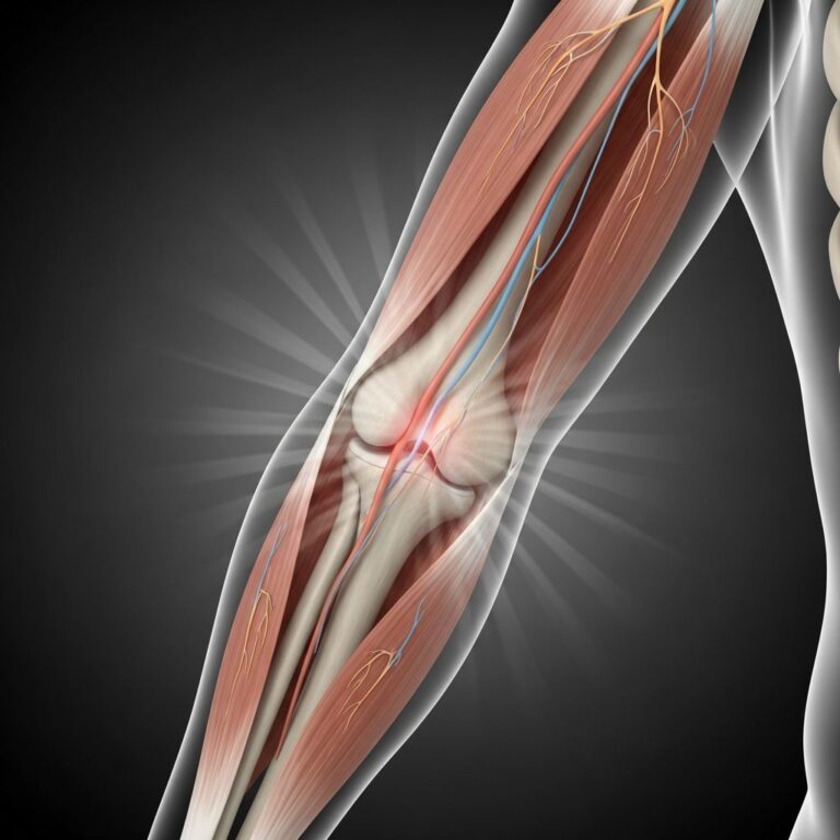
MRI is the gold standard for evaluating degenerative changes in the spine, including disc herniation, bulging discs, and foraminal stenosis. T2-weighted sequences show the hydration status of intervertebral discs, with degenerated discs appearing darker due to loss of water content. The relationships between degenerative changes and neural compression can be assessed in multiple planes, helping guide treatment decisions.
Spinal Cord Injury and Myelopathy
In acute spinal cord injury, MRI can detect cord edema, hemorrhage, and structural disruption, providing prognostic information. For chronic myelopathy, whether from cervical spondylosis, syrinx formation, or other causes, MRI demonstrates both the structural abnormalities and their impact on the spinal cord.
Infection and Inflammation
Spinal infections, including osteomyelitis, discitis, and epidural abscess, can be identified with MRI. T1-weighted images with contrast enhancement help delineate areas of infection, while STIR sequences show associated edema. Similarly, inflammatory conditions affecting the spine can be characterized and monitored.
Preparing for Brain and Spine MRI
Pre-Scan Screening
Before undergoing MRI, patients must complete a thorough screening questionnaire to identify contraindications and safety concerns. The strong magnetic field can interact with ferromagnetic materials, making it essential to identify any metallic implants.
Contraindications and Precautions
- Certain cardiac pacemakers and implantable cardioverter-defibrillators (ICDs)
- Ferromagnetic aneurysm clips
- Metallic foreign bodies in the eyes
- Some neurostimulators and cochlear implants
- Certain metallic tattoos or permanent makeup
Many modern implants are MRI-compatible, but specific models and field strengths must be verified. Patients should inform their healthcare provider about any medical devices, implants, or metallic objects before scheduling.
What to Expect During the Procedure
MRI procedures typically last 30 to 60 minutes, depending on the specific protocol and number of sequences required. Patients are positioned on a sliding table that moves into the scanner bore. The scanner makes loud knocking and humming sounds as the magnetic field is switched on and off. Earplugs or headphones are provided to minimize noise exposure. A call button allows patients to communicate with the technician if needed.
Remaining still during scanning is crucial for image quality. Even small movements can degrade image resolution and may require repeating sequences, extending scan duration. For patients with claustrophobia or anxiety, sedation may be available, though this requires discussion with the imaging facility beforehand.
Advanced MRI Techniques
Diffusion Tensor Imaging (DTI)
DTI measures the directional movement of water molecules along white matter tracts, allowing visualization and quantification of neural connectivity. This technique is valuable for pre-surgical planning, assessing white matter integrity in neurodegenerative diseases, and researching brain organization.
Functional MRI (fMRI)
Functional MRI detects changes in blood oxygenation associated with neural activity, allowing visualization of brain regions activated during specific tasks. This technique is used for research and for pre-operative mapping of eloquent cortex in tumor cases.
Magnetic Resonance Spectroscopy (MRS)
MRS analyzes the chemical composition of brain tissue by detecting signals from specific metabolites. This can help characterize tumors, detect metabolic abnormalities in neurodegenerative diseases, and assess tissue viability.
Quantitative Susceptibility Mapping (QSM)
QSM is an advanced post-processing technique derived from SWI sequences that quantifies local magnetic field disturbances caused by iron and other paramagnetic substances. This allows objective measurement of iron content in specific brain regions, valuable for assessing Parkinson’s disease, Alzheimer’s disease, and other conditions associated with abnormal iron accumulation.
Safety Considerations
Magnetic Field Safety
While the static magnetic field itself is generally considered safe, the rapidly changing magnetic fields and radiofrequency pulses can pose risks under certain circumstances. Patients with implanted devices require careful evaluation before proceeding with MRI.
Gadolinium Contrast Agents
Contrast-enhanced MRI uses gadolinium-based agents to enhance visualization of certain pathologies. While generally safe, gadolinium is contraindicated in patients with severe renal impairment due to the risk of nephrogenic systemic fibrosis. The radiologist should be informed of any kidney disease before contrast administration.
Pregnancy Considerations
MRI is generally considered safe in pregnancy, particularly after the first trimester, as it does not use ionizing radiation. However, contrast agents should typically be avoided unless medically necessary. Pregnant patients should discuss MRI risks and benefits with their healthcare provider.
Comparing MRI with Other Imaging Modalities
| Imaging Modality | Radiation Exposure | Soft Tissue Contrast | Speed | Cost | Best Use Cases |
|---|---|---|---|---|---|
| MRI | None | Excellent | 30-60 minutes | High | Brain/spine pathology, soft tissue detail |
| CT | Yes | Good | 1-5 minutes | Moderate | Acute trauma, bone detail, quick screening |
| X-ray | Yes | Poor | Seconds | Low | Bone fractures, basic screening |
| Ultrasound | None | Fair | 10-30 minutes | Low | Real-time imaging, blood flow assessment |
Frequently Asked Questions
Q: Is MRI safe for patients with metal implants?
A: It depends on the type of implant. Many modern implants are MRI-compatible, but older ferromagnetic devices may contraindicate MRI. Always inform your healthcare provider about any implants so proper screening can be performed.
Q: How long does a brain or spine MRI take?
A: Most brain and spine MRI studies take between 30 to 60 minutes, depending on the specific protocols needed and whether contrast is administered.
Q: Can I eat or drink before an MRI?
A: Generally, you can eat and drink normally unless sedation is planned. If sedation is needed, fasting requirements will be provided in your pre-procedure instructions.
Q: Will I feel anything during the MRI?
A: You will hear loud knocking and humming sounds but should not feel pain. Some patients experience mild warmth or a tingling sensation, which is normal.
Q: Is MRI better than CT for brain imaging?
A: MRI provides superior soft tissue contrast and is better for evaluating most brain pathology. However, CT is faster and better for detecting acute bleeding in emergency situations.
Q: Can patients with claustrophobia undergo MRI?
A: Yes. Many facilities offer open MRI scanners or allow sedation for claustrophobic patients. Discuss your concerns with the imaging facility in advance.
Q: How soon will I get my MRI results?
A: A radiologist typically reviews the images and provides a report to your physician within 24 to 48 hours, though urgent cases are prioritized.
Q: Is contrast always necessary for brain and spine MRI?
A: No. Contrast is not always required and depends on the clinical question. Your radiologist will determine whether contrast is needed for your specific examination.
References
- Susceptibility weighted imaging: Clinical applications and future perspectives — National Center for Biotechnology Information. 2018. https://pmc.ncbi.nlm.nih.gov/articles/PMC5971274/
- Magnetic Resonance Imaging of the Brain and Spine — Johns Hopkins University. 1988. https://pure.johnshopkins.edu/en/publications/magnetic-resonance-imaging-of-the-brain-and-spine-4
- MRI Physics: Magnetic Resonance and Spin Echo Sequences — Johns Hopkins Medicine. 2022. Educational resource on fundamental MRI physics principles and clinical applications.
Similar Articles


Read full bio of Sneha Tete