Mycology Guide To Diagnosing Fungal Skin Infections
Comprehensive guide to the study of fungi causing skin, nail, and hair infections, including diagnosis and classification.

Skin scrapings and nail clippings are essential specimens taken to establish or confirm the diagnosis of a fungal infection through microscopy and culture, known as mycology. Superficial fungal infections primarily affect the outer layers of the skin, nails, and hair, caused predominantly by dermatophytes from the genera Trichophyton, Microsporum, and Epidermophyton.
What is mycology?
Mycology is the scientific study of fungi, a diverse kingdom of eukaryotic organisms distinct from plants, animals, and bacteria. In dermatology, mycology focuses on fungi pathogenic to human skin, nails, and hair. Fungi can be yeasts, moulds, or dimorphic, reproducing asexually or sexually via spores. Mycologists classify them by microscopic appearance, culture growth, and reproductive methods.
Growing fungi form branched filaments called hyphae, collectively known as the mycelium. Hyphae may be septate (compartmentalized by cross-walls) or aseptate. Yeasts are unicellular fungi that bud to reproduce, forming clusters of round or oval cells.
Superficial fungal infections
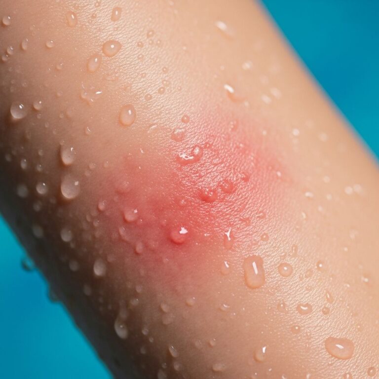
These infections are confined to the epidermis, hair, and nails. Dermatophyte infections (tinea or ringworm) are named by affected site: tinea pedis (feet), tinea corporis (body), tinea capitis (scalp), etc. Dermatophytes require keratin for nutrition and are classified as anthropophilic (human-adapted), zoophilic (animal-adapted), or geophilic (soil-adapted).
- Anthropophilic: Trichophyton rubrum, T. tonsurans – mild inflammation, chronic infections.
- Zoophilic: Microsporum canis, T. verrucosum – inflammatory, acute.
- Geophilic: Microsporum gypseum – highly inflammatory.
Yeasts like Candida species (e.g., C. albicans) and Malassezia cause infections in moist areas or follicles. Candida is a commensal turning pathogenic with host changes; Malassezia causes pityriasis versicolor and folliculitis.
Diagnosis of superficial fungal infections
Diagnosis relies on clinical suspicion confirmed by laboratory tests. Collect skin scrapings from active edges, nail clippings from proximal undersurface, or hair pluckings.
Microscopy
Potassium hydroxide (KOH) 10-20% dissolves keratin, revealing hyphae, spores, or yeast cells under light microscopy. Add calcofluor white or periodic acid-Schiff (PAS) for fluorescence. Dermatophytes show branching septate hyphae; yeasts appear as budding cells or pseudohyphae.
Culture
Inoculate on Sabouraud dextrose agar (SDA) with cycloheximide and chloramphenicol. Incubate at 25-30°C for 1-4 weeks. Observe colony morphology (color, texture, pigment), then microscopic features like macroconidia for genus identification.
| Fungus Type | Microscopy Features | Culture Characteristics |
|---|---|---|
| Dermatophytes | Septate hyphae, arthroconidia | Velvety/ powdery colonies, slow growth |
| Candida | Pseudohyphae, blastospores | Creamy white, yeast-like |
| Malassezia | Spherical yeasts, short hyphae | Lipophilic, requires oils |
Implications of mycology results
Positive microscopy indicates infection but not species; culture identifies the pathogen for targeted therapy. Negative results do not exclude infection if sampling is poor. Resistant strains (e.g., T. indotineae) require susceptibility testing.
In histology from biopsies, Grocott-Gomori methenamine silver (GMS) or PAS stains highlight fungi in tissue, crucial for atypical presentations like Majocchi’s granuloma.
Other fungal infections
Subcutaneous mycoses (e.g., sporotrichosis, chromoblastomycosis) arise from traumatic inoculation of environmental moulds into dermis. Diagnosed by culture and histology showing muriform cells or asteroid bodies.
Systemic mycoses (e.g., histoplasmosis, cryptococcosis) disseminate to skin in immunocompromised patients. Skin lesions mimic other conditions; diagnosis involves blood cultures, serology, and PCR alongside histopathology.
Collection of specimens
Clean site with alcohol. Scrape scaling edge with scalpel #15 into sterile container. For nails, clip proximal; hair, pluck with forceps. Transport promptly to lab.
- Skin: Active border scales.
- Nails: Clippings + subungual debris.
- Hair: Fluorescing or broken shafts.
Frequently Asked Questions (FAQs)
Q: What does a positive KOH mount show?
A: Branching hyphae or yeast forms confirm fungal elements, guiding empirical treatment.
Q: How long does fungal culture take?
A: 1-4 weeks; preliminary reports at 7-14 days.
Q: Can superficial fungi cause deep infections?
A: Rarely, via follicles (Majocchi’s granuloma); biopsy needed.
Q: What if culture is negative but microscopy positive?
A: Treat based on microscopy; repeat sample or consider PCR.
Q: Are all ringworm fungi dermatophytes?
A: No, some tinea-like infections from yeasts or non-dermatophytes.
This comprehensive overview equips clinicians to accurately diagnose and manage fungal infections through mycology. Early, precise identification prevents spread and resistance.
References
- Mycology – Fungal skin infections — DermNet NZ. 2023. https://dermnetnz.org/cme/fungal-infections/mycology
- Dermatopathology and the Diagnosis of Fungal Infections — PMC (NIH). 2023-06-01. https://pmc.ncbi.nlm.nih.gov/articles/PMC10282148/
- Introduction to fungal infections — DermNet NZ. 2023. https://dermnetnz.org/topics/introduction-to-fungal-infections
- Collecting specimens for the investigation of fungal infections — bpac.org.nz. 2011-03-01. https://bpac.org.nz/BT/2011/March/fungal-infections.aspx
- Mycology of dermatophyte infections — DermNet NZ. 2023. https://dermnetnz.org/topics/mycology-of-dermatophyte-infections
Similar Articles


Read full bio of medha deb