Mycosis Fungoides: Diagnosis, Staging, And Treatment Guide
Comprehensive guide to mycosis fungoides: symptoms, stages, diagnosis, and advanced treatment options for this rare cutaneous T-cell lymphoma.

Mycosis fungoides (MF) is the most common form of
cutaneous T-cell lymphoma
(CTCL), a group of non-Hodgkin lymphomas originating from T-lymphocytes in the skin. It typically presents with persistent, itchy patches that slowly evolve over years, affecting primarily adults over 50 years old.What is mycosis fungoides?
Mycosis fungoides is a rare, chronic
primary T-cell lymphoma
confined initially to the epidermis, with neoplastic T-cells exhibiting a mature helper T-cell (CD4+) immunophenotype. Despite its name suggesting fungal origin (‘mycosis’ means fungal, ‘fungoides’ mushroom-like), it is not caused by fungi but named for tumor resemblance to mushrooms. It accounts for about 50% of CTCL cases and has an incidence of 6.4 per million, more common in males and African Americans.The disease progresses slowly through patch, plaque, and tumor stages, with potential extracutaneous spread in advanced cases. Early-stage MF (<10% skin involvement) has a favorable prognosis, with 5-year survival near 95%.
Who gets mycosis fungoides?
- Primarily adults aged 50–70 years.
- Male predominance (2:1 ratio).
- Higher incidence in Black individuals.
- Rare in children (<2% of cases).
- No strong genetic predisposition identified, though associations with human retroviruses debated.
What causes mycosis fungoides?
The exact cause remains unknown. It arises from clonal proliferation of malignant CD4+ T-cells with a skin-homing phenotype (CLA+, CCR4+). Potential triggers include:
- Chronic antigenic stimulation (e.g., infections, autoimmunity).
- Viral factors (HTLV-1 debated, not confirmed).
- Environmental exposures (pesticides, industrial chemicals).
- Genetic mutations in TCR, FAS, TP53 genes.
No infectious agent definitively implicated; not contagious.
What are the clinical features of mycosis fungoides?
Early lesions mimic benign dermatoses. Progression is indolent, spanning 5–20 years from patches to tumors.
Patch stage (IA–IB)
Superficial, erythematous, scaly patches, often pruritic, on sun-protected sites (buttocks, thighs, breasts). Geographic borders, fine scale; may persist >6 months.
Plaque stage (IB–IIA)
Indurated, elevated plaques, larger, more symptomatic. Poikiloderma (mottled pigmentation, atrophy, telangiectasia) common.
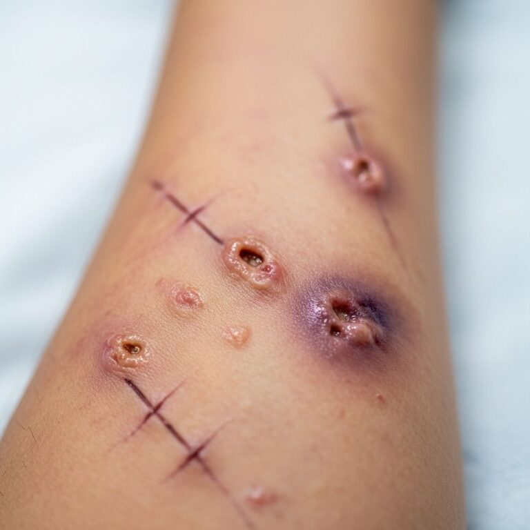
Tumour stage (IIB)
Circumscribed, dome-shaped, ulcerated tumors. Rapid growth possible.
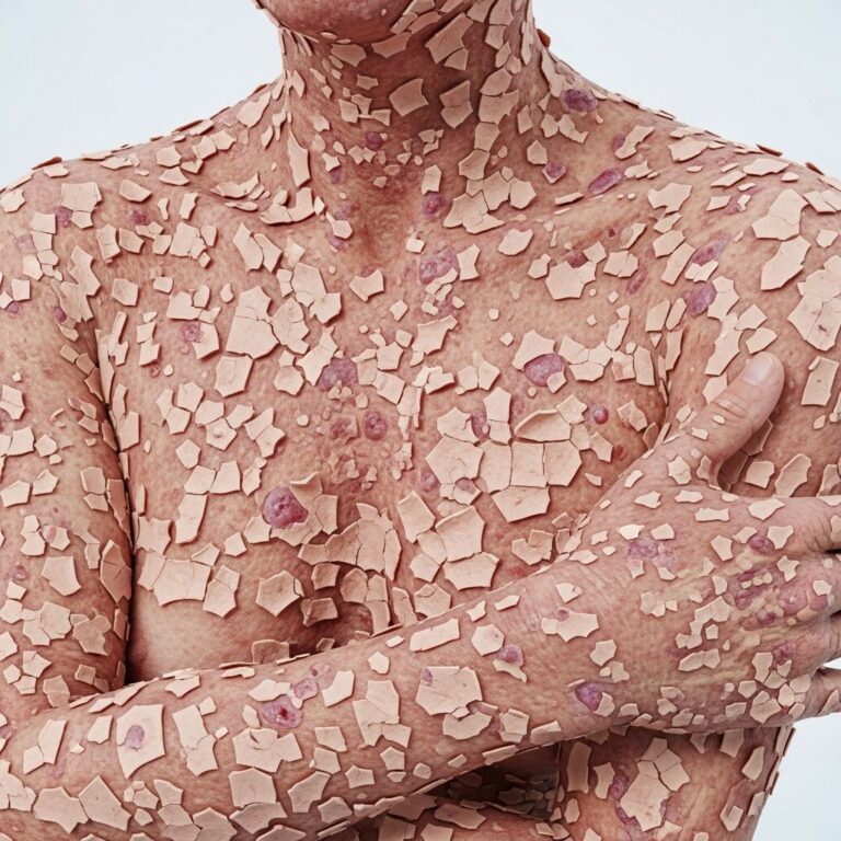
Erythroderma (IIIB–IV)
Generalized redness, scaling; may overlap Sézary syndrome (with circulating atypical cells).
Other features
- Pruritus (universal, severe).
- Alopecia, nail dystrophy.
- Lymphadenopathy (firm, rubbery nodes).
- Mucosal involvement rare.
How is mycosis fungoides diagnosed?
Diagnosis requires clinicopathological correlation: persistent lesions unresponsive to standard therapies.
- Skin biopsy: Dense lymphocytic infiltrate, epidermotropism (atypical lymphocytes in epidermis), Pautrier microabscesses. Immunohistochemistry: CD4+, CD3+, often CD7-, CD62L- loss.
- T-cell receptor (TCR) analysis: Clonal rearrangement confirms lymphoma.
- Staging workup: Blood (Sézary cells, flow cytometry), nodes (excisional biopsy), imaging (CT/PET-CT for advanced disease).
What is the staging of mycosis fungoides?
TNM-B system (ISCL/EORTC):
| Stage | Skin (T) | Nodes (N) | Visceral (M) | Blood (B) | Prognosis |
|---|---|---|---|---|---|
| IA | T1: <10% patches/plaques | N0 | M0 | B0 | Excellent (>95% 10-yr survival) |
| IB | T2: >10% patches/plaques | N0 | M0 | B0 | Good |
| IIA | T1–2 | N1 (dermatopathic) | M0 | B0–1 | Moderate |
| IIB | T3: Tumors | N0–1 | M0 | B0–1 | Fair |
| III | T4: Erythroderma | N0–2 | M0 | B0–2 | Moderate |
| IVA1 | T1–4 | N0–2 | M0 | B2 | Poor |
| IVA2 | T1–4 | N3: Nodal lymphoma | M0 | B0–2 | Poor |
| IVB | T1–4 | N0–3 | M1 | B0–2 | Very poor |
Early stages (I–IIA): skin-limited, >90% 10-year survival. Advanced (III–IV): 40–60% 5-year survival.
What is the treatment for mycosis fungoides?
Treatment is stage-directed, palliative in advanced disease. Goals: symptom control, quality of life.
Skin-directed therapies (early stage IA–IIA)
- Topical corticosteroids: First-line for limited patches/plaques (clobetasol 0.05%). Response 60–90%.
- Topical chemotherapy: Mechlorethamine (nitrogen mustard) gel, FDA-approved for IA–IB. Applied daily; 70–80% response.
- Topical retinoids: Bexarotene 1% gel.
- Phototherapy: Narrowband UVB (NB-UVB) for thin patches; PUVA (psoralen+UVA) for plaques/tumors. CR rates 80–95% early disease.
- Localized radiation: Electron beam for unilesional/tumors.
Systemic therapies (advanced/refractory)
- Retinoids: Bexarotene oral (FDA-approved CTCL), 45% response.
- Interferon-alpha: Subcutaneous, 50–70% response early disease.
- Low-dose methotrexate: <50 mg/week, ORR 33%.
- Histone deacetylase inhibitors: Vorinostat, romidepsin (FDA-approved refractory CTCL).
- Monoclonal antibodies: Mogamulizumab (Poteligeo, FDA 2018 for relapsed MF).
- Checkpoint inhibitors: Pembrolizumab (trials).
- Chemotherapy: Limited role (pegylated liposomal doxorubicin, gemcitabine).
- Stem cell transplant: Allogeneic for young advanced patients.
Extracorporeal photopheresis (ECP)
For erythrodermic MF/Sézary; draws/treats/reinfuses blood with 8-methoxypsoralen+UVA. Improves survival.
What is the outcome for mycosis fungoides?
Prognosis stage-dependent:
- IA: >95% 30-year survival.
- IB: 85–90% 10-year.
- IIB–III: 50–65% 5-year.
- IV: <30% 5-year.
Adverse factors: thickness, visceral involvement, high LDH. Disease-specific survival better than all-cause due to comorbidities.
How can mycosis fungoides be prevented?
No known prevention; early recognition key. Avoid irritants, treat infections promptly.
Frequently Asked Questions
What is the difference between mycosis fungoides and Sézary syndrome?
MF is skin-predominant; Sézary syndrome is leukemic variant with >1000/μL Sézary cells, generalized erythroderma.
Is mycosis fungoides curable?
Curative in early stage IA with skin-directed therapy; advanced disease managed palliatively.
Does mycosis fungoides spread?
<10% early cases disseminate; risk increases with stage.
How long can you live with mycosis fungoides?
Median survival 10–35 years; excellent for early stages.
Is mycosis fungoides hereditary?
No familial cases reported; sporadic.
References
- Mycosis Fungoides – Symptoms, Causes, Treatment — NORD (rarediseases.org). 2023. https://rarediseases.org/rare-diseases/mycosis-fungoides/
- Mycosis fungoides: Pictures and symptoms by stage — Medical News Today. 2023-05-26. https://www.medicalnewstoday.com/articles/mycosis-fungoides
- How We Treat Mycosis Fungoides and Sézary Syndrome — PMC (NCBI). 2022-08-11. https://pmc.ncbi.nlm.nih.gov/articles/PMC9364355/
- Mycosis Fungoides (Including Sézary Syndrome) Treatment – NCI — National Cancer Institute (cancer.gov). 2025. https://www.cancer.gov/types/lymphoma/patient/mycosis-fungoides-treatment-pdq
- Mycosis Fungoides — Cutaneous Lymphoma Foundation. 2023. https://www.clfoundation.org/mycosis-fungoides
- How I treat mycosis fungoides and Sézary syndrome — Blood (ASH Publications). 2016-06-02. https://ashpublications.org/blood/article/127/25/3142/35196/How-I-treat-mycosis-fungoides-and-Sezary-syndrome
Similar Articles


Read full bio of medha deb