Myelin Sheath: What It Is, Purpose & Function
Understanding myelin sheath: the protective coating that speeds up nerve signals.

Understanding the Myelin Sheath
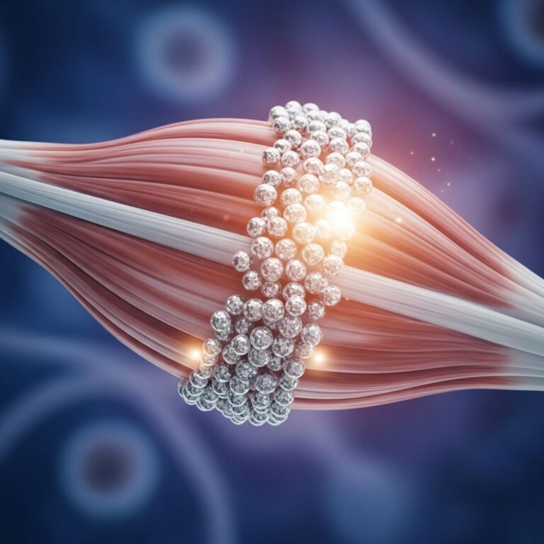
The myelin sheath is a protective membrane that wraps around parts of certain nerve cells, playing a crucial role in how efficiently your nervous system communicates. This lipid-rich, white substance acts as an insulating layer surrounding the axons of neurons throughout both the central and peripheral nervous systems. Without the myelin sheath, your nerve signals would travel much more slowly, and your brain and body wouldn’t be able to communicate as effectively. Understanding this essential component of your nervous system helps illuminate how your body processes information, coordinates movements, and maintains overall neurological health.
What Is the Myelin Sheath?
The myelin sheath is a greatly extended and modified plasma membrane that wraps around a nerve axon in a spiral fashion, forming multiple concentric layers of insulation. Imagine wrapping a sleeping bag around a cylindrical object repeatedly—this visual representation captures how the myelin sheath forms around the axon. The sheath itself consists of approximately 100 layers of tightly packed plasma membrane, each layer meticulously arranged to provide maximum insulation and protection.
This specialized coating is not uniform throughout the entire axon. Instead, the myelin sheath covers segments of the axon with small gaps between these segments called nodes of Ranvier. These nodes are crucial unmyelinated sections where the axon membrane is exposed, allowing for the unique conduction process that makes nerve signal transmission so rapid and efficient.
Where Is the Myelin Sheath Found?
The myelin sheath is found in both the central nervous system (brain and spinal cord) and the peripheral nervous system (nerves extending throughout the body). However, not all axons are myelinated. Some axons remain unmyelinated, meaning they lack this protective coating. The presence or absence of myelin significantly affects how quickly and efficiently nerve signals travel.
Myelinated axons are typically found in pathways where speed is essential—such as motor nerves that control muscle movement and sensory nerves that transmit pain, temperature, and touch sensations. Unmyelinated axons are more common in functions that don’t require rapid signal transmission, such as in some autonomic nervous system pathways that regulate internal body functions like heart rate and digestion.
How Is the Myelin Sheath Created?
The myelin sheath is produced by specialized glial cells that differ between the central and peripheral nervous systems. Understanding how these cells create the myelin sheath reveals the remarkable precision of your nervous system.
Production in the Peripheral Nervous System
In the peripheral nervous system, Schwann cells are responsible for creating the myelin sheath. During fetal development, Schwann cells begin the myelination process by wrapping their lipid-rich membrane around axons multiple times. As the wrapping continues, the nucleus and cytoplasm of the Schwann cell are gradually squeezed out toward the outer layer. Once myelination is complete, the Schwann cell’s nucleus and cytoplasm reside in the outermost layer, creating a structure called the neurilemma. This arrangement means that each Schwann cell can only myelinate one axon segment at a time.
Production in the Central Nervous System
In the central nervous system, oligodendrocytes perform the myelination function. Unlike Schwann cells, a single oligodendrocyte has approximately 15 flat, broad, arm-like processes extending from its cell body, allowing it to myelinate multiple axons simultaneously. This efficiency is remarkable—one oligodendrocyte can myelinate up to 40 or more separate axons at the same time. The oligodendrocyte can generate all the necessary myelin proteins and lipids in just about 5 hours, demonstrating the efficiency of this biological process.
In the central nervous system, the cell body and nucleus of oligodendrocytes remain separate from the myelin sheath, meaning there is no neurilemma present as there is with Schwann cells. Instead, a small glial tongue of cytoplasm extends through slender processes to connect with the plasma membrane of the oligodendroglial cell.
The Structure and Composition of Myelin
The myelin sheath has a remarkably organized structure that contributes to its insulating properties. The tight, layer-by-layer packing of the myelin sheath creates characteristic periodic morphological features visible under electron microscopy.
Key Structural Features
The myelin sheath contains two characteristic repeating structures: major dense lines and intraperiod lines. The major dense lines are approximately 2 to 3 nanometers wide and are formed by closely condensed intracellular (cytoplasmic) surfaces between the inner membranes of lipid bilayers. These lines represent areas where the cytoplasmic sides of the membrane are pressed together, creating an extremely tight seal. The intraperiod lines represent the extracellular surface areas between membranes.
At the edges of each myelin segment, individual myelin lamellae attach to the axon as cytoplasm-containing terminal loops. These loop-shaped terminations are called lateral loops and form membrane complexes with the axolemma (the axon’s membrane) called transverse bands. These transverse bands are helical structures that seal the myelin to the axon while still providing, through small spaces between them, a pathway from the extracellular space to the periaxonal space.
Molecular Composition
The myelin sheath is primarily composed of lipids and proteins, with lipids comprising about 70-80% of the dry weight of myelin. This high lipid content gives myelin its characteristic white appearance and its excellent insulating properties. Myelin basic protein (MBP) is essential for the compaction of the two adjacent cytoplasmic membrane surfaces into the major dense line of myelin.
Other important proteins include myelin-associated glycoprotein (MAG), which is localized on the inner membrane of the myelin sheath and interacts with axonal membrane proteins to attach the myelin sheath to the axon. The composition of myelin varies slightly between the central and peripheral nervous systems, reflecting their different functional requirements and cellular origins.
Primary Functions of the Myelin Sheath
The myelin sheath serves several critical functions that make it essential for proper nervous system function. While insulation is its most well-known role, recent research has revealed additional important functions.
Rapid Signal Conduction
The primary function of the myelin sheath is to insulate the axon and increase the velocity of action potential propagation. Myelin has properties of low capacitance and high electrical resistance, which means it can act as an excellent electrical insulator. This insulation allows nerve signals to travel much faster than they would in unmyelinated axons. Myelinated axons can conduct action potentials at speeds up to 120 meters per second, while unmyelinated axons conduct signals at only about 1 meter per second.
Saltatory Conduction
One of the most efficient mechanisms enabled by myelin is saltatory conduction. Instead of the action potential traveling continuously along the entire axon, it “jumps” from one node of Ranvier to the next, dramatically increasing conduction speed. This jumping motion is possible because the myelin sheath insulates the axon between nodes, preventing the depolarization of the membrane in these regions. The voltage-gated sodium channels are clustered at the nodes of Ranvier, where they can be activated to regenerate the action potential.
Signal Containment and Protection
The myelin sheath prevents electrical signals from leaking out of the axon, preserving signal strength and direction. This containment is essential for accurate and efficient neural communication. The tight seals of myelin with their apposing lamellae are crucial for these insulating properties, preventing current leakage and allowing for efficient and fast nerve conduction.
Ion Regulation
Myelin helps control sodium and potassium flow, which is essential for nerve impulses. Paranodal junctions—specialized contact points between myelin and axons—help keep signal-related proteins in the right places. These junctions ensure that ions are properly separated, preventing signal disruption. During signal transmission, potassium levels rise around the axon, and myelin contains channels that help remove this excess potassium, while nearby astrocytes absorb and transport it out of the nervous system.
Energy Efficiency
By concentrating ion channels at specific points (the nodes of Ranvier) and reducing the need for continuous ion exchange along the entire axon, myelin minimizes the energy required for repeated signal transmission. This energy efficiency is crucial, as the nervous system is one of the most metabolically demanding systems in the body. The myelin sheath also may provide trophic support to the underlying axon, with some evidence suggesting that oligodendrocytes can supply lactate to axons for ATP generation.
Axon Sculpting
Beyond its insulating role, the myelinating cell “sculpts” the underlying axon by promoting the phosphorylation of neurofilaments, thus increasing the diameter or thickness of the axon at the internodal regions. This sculpting function helps optimize the axon’s properties for efficient signal conduction.
The G-Ratio: Optimizing Myelin Thickness
The g-ratio—the ratio of axon diameter to the outer diameter of the myelinated axon (including myelin)—is an important concept in understanding myelin function. Oligodendrocytes myelinate different axons to variable extents depending on axon diameter to maintain an optimal g-ratio. This means that larger axons receive thicker myelin coatings compared to smaller axons. This optimization ensures that each axon achieves maximum conduction velocity for its particular size, demonstrating the remarkable precision of the nervous system.
Myelin Development and Maturation
Myelination is not complete at birth. In the brain, myelination continues throughout development, with mature myelination typically being achieved at approximately 2 years of age. However, some aspects of myelination continue into early adulthood, particularly in the prefrontal cortex and other regions involved in complex cognitive functions. This developmental timeline is important for understanding childhood development and why certain abilities emerge at specific ages.
Clinical Significance and Demyelinating Diseases
When the myelin sheath is damaged or destroyed, nerve signal transmission becomes impaired, leading to various neurological symptoms. Demyelinating diseases such as multiple sclerosis involve the loss of myelin, which disrupts normal nerve function. Understanding the structure and function of the myelin sheath is crucial for developing treatments for these conditions. Mutations to genes coding for myelin proteins, such as the MAG gene, are implicated in demyelination diseases.
Frequently Asked Questions
Q: What exactly is the myelin sheath?
A: The myelin sheath is a protective, lipid-rich membrane that wraps around nerve axons in multiple layers, acting as an insulator to speed up electrical signal transmission through the nervous system.
Q: How fast do signals travel in myelinated versus unmyelinated axons?
A: Myelinated axons conduct action potentials at speeds up to 120 meters per second, while unmyelinated axons conduct signals at approximately 1 meter per second—a dramatic difference in speed.
Q: Which cells produce the myelin sheath?
A: In the peripheral nervous system, Schwann cells produce myelin, with each cell myelinating one axon segment. In the central nervous system, oligodendrocytes produce myelin and can simultaneously myelinate up to 40 or more axon segments.
Q: What is saltatory conduction?
A: Saltatory conduction is the process where action potentials jump from one node of Ranvier to the next along a myelinated axon, allowing for rapid signal transmission.
Q: How does myelin contribute to energy efficiency?
A: Myelin concentrates ion channels at the nodes of Ranvier, reducing the need for continuous ion exchange along the entire axon. This targeted approach minimizes the energy required for repeated signal transmission.
Q: At what age is myelination complete in the brain?
A: Brain myelination is typically mature by approximately 2 years of age, though some aspects of myelination continue into early adulthood.
Q: What are demyelinating diseases?
A: Demyelinating diseases, such as multiple sclerosis, involve damage to or loss of the myelin sheath, which disrupts normal nerve function and causes various neurological symptoms.
Q: What is the g-ratio?
A: The g-ratio is the ratio of axon diameter to the outer diameter of the myelinated axon (including myelin). Oligodendrocytes optimize this ratio to ensure maximum conduction velocity for each axon size.
References
- Myelin Sheath: What It Is, Purpose & Function — Cleveland Clinic. 2024. https://my.clevelandclinic.org/health/body/22974-myelin-sheath
- The Myelin Sheath – Basic Neurochemistry — National Center for Biotechnology Information (NCBI). https://www.ncbi.nlm.nih.gov/books/NBK27954/
- Myelin Sheath — Simply Psychology. https://www.simplypsychology.org/myelin-sheath.html
- Myelin in the Central Nervous System: Structure, Function, and Pathology — Physiological Reviews, American Physiological Society. 2018. https://journals.physiology.org/doi/abs/10.1152/physrev.00031.2018
- Overview of Myelin, Major Myelin Lipids, and Myelin-Associated Proteins — Frontiers in Chemistry. 2022. https://www.frontiersin.org/journals/chemistry/articles/10.3389/fchem.2022.1041961/full
- Myelin: Structure, Function, and Associated Disorders — Kenhub. https://www.kenhub.com/en/library/anatomy/the-myelin-sheath-and-myelination
Similar Articles


Read full bio of medha deb