Naevus Lipomatosus Superficialis Pathology
Comprehensive pathology guide to naevus lipomatosus superficialis, a benign cutaneous hamartoma with ectopic dermal adipocytes.

Introduction
Naevus lipomatosus superficialis (NLS) represents a rare benign cutaneous hamartoma characterized by the ectopic accumulation of mature adipocytes within the dermis, distinct from the typical subcutaneous location of fat cells. This developmental malformation manifests as clusters of soft, yellowish papules or nodules, primarily affecting the pelvic girdle region. First described by Hoffman and Zurhelle in 1921, NLS is classified into two main variants: the classical (multiple) type, also known as Hoffmann-Zurhelle type, and the solitary type. The classical form typically presents in infancy or early childhood with linear or zosteriform arrangements of lesions, while the solitary variant may appear later in life as an isolated nodule anywhere on the body. Although generally asymptomatic, these lesions pose primarily cosmetic concerns, with rare associations to ulceration or necrosis. Understanding the pathology of NLS is crucial for accurate diagnosis and differentiation from other dermal proliferations.
Demographics
NLS exhibits no significant sex predilection, occurring equally in males and females. The classical multiple type predominantly emerges at birth or within the first three decades of life, often noticed in infancy as congenital birthmarks. Lesions in this variant favor the lower trunk, including the buttocks, lumbar region, sacrum, pelvic girdle, and upper thighs, frequently displaying a segmental or zosteriform distribution along Blaschko’s lines or nerve distributions. In contrast, the solitary type may present later, even in adulthood, and can occur on diverse sites such as the scalp, face, neck, extremities, or trunk without a specific pattern. Rare cases of generalized involvement have been documented, though these are exceptional. Pediatric presentations are more common for the classical form, with growth stabilizing or progressing slowly into adulthood. The condition’s rarity underscores the importance of histopathological confirmation in suspected cases.
Causes
The precise etiology of naevus lipomatosus superficialis remains unknown, though it is widely regarded as a developmental anomaly arising from mesenchymal dysplasia during embryogenesis. Theories propose ectopic differentiation of adipogenic precursor cells within the dermis, potentially influenced by local intrauterine pressures in the pelvic girdle, where persistent fat pad thickness may predispose to disordered fat cell migration. Some researchers classify NLS as a connective tissue naevus due to associated alterations in collagen, elastic fibers, fibroblasts, and occasional pilar or sebaceous elements, suggesting a broader hamartomatous process. Genetic factors are not well-defined, with no consistent chromosomal abnormalities identified, though mosaic mutations along developmental lines could explain segmental patterns. Unlike true lipomas, NLS lacks capsule formation and shows no connection to subcutaneous fat, supporting its hamartomatous rather than neoplastic nature. Environmental triggers or trauma are not implicated, reinforcing its congenital basis.
Clinical features
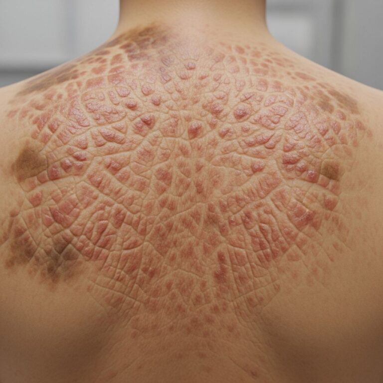
Clinically, the classical type of NLS presents as multiple, soft, dome-shaped or pedunculated papules and nodules, ranging from 2-30 mm in diameter, clustered into plaques with a smooth, wrinkled, cerebriform, or rarely warty surface. These lesions are typically skin-colored to yellowish, non-tender, and mobile over deeper structures, often arranged linearly, zosteriform, or segmentally along skin folds or Blaschko’s lines on the lower back, buttocks, thighs, or pelvic girdle. Hairiness, comedo-like plugs, or peau d’orange texture may occur. The solitary variant appears as a single, asymptomatic, rubbery subcutaneous swelling, polypoid or sessile, measurable up to several centimeters, capable of arising on any body site including axillae, extremities, or head and neck. Lesions grow slowly, remaining stable or enlarging gradually, without spontaneous regression. Patients rarely report symptoms beyond cosmetic dissatisfaction.
- Sites of predilection: Pelvic girdle (80-90% of classical cases), buttocks, lumbar/sacral area, upper thighs.
- Rare locations: Scalp, face, neck, shoulders, thorax, abdomen, genitalia, extremities, soles.
- Morphology: Soft, yellow-orange papules/nodules/plaques; pedunculated or sessile; smooth/cerebriform/wrinkled surface.
- Distribution: Zosteriform, linear, segmental in classical type; solitary in isolated form.
Complications
Naevus lipomatosus superficialis is predominantly benign with no malignant potential or systemic associations. Complications are infrequent but include rare ulceration, necrosis, foul-smelling discharge, or pain, possibly due to compression of dermal vessels by ectopic adipocytes or external trauma from frictional sites like the pelvic girdle. Secondary infection may supervene in ulcerated lesions. Cosmetic disfigurement remains the primary issue, particularly with progressive coalescence into larger plaques. No hereditary patterns or syndromic links (e.g., to Goltz syndrome) are established, though isolated co-occurrences with naevus sebaceous have been reported. Growth acceleration during puberty or pregnancy is anecdotal. Overall, morbidity is low, focused on aesthetic impact.
Diagnosis
Definitive diagnosis of NLS relies on histopathological examination of a biopsy or excisional specimen. Key microscopic features include clusters or sheets of mature adipocytes scattered ectopically in the reticular dermis, comprising 10-50% of the lesion volume, without encapsulation or subcutaneous extension. Adipocytes appear normal, often clustered around blood vessels, eccrine glands, or collagen bundles, accompanied by variable fibroplasia, increased fibroblasts, thickened collagen, and elastic fibers. The epidermis may show mild acanthosis, hyperkeratosis, basal hyperpigmentation, or elongated rete ridges. Adnexal structures can exhibit perifollicular fibrosis or reduction. No atypia, mitoses, or lipoblasts are present, distinguishing it from liposarcoma. Dermoscopy may reveal yellowish structureless areas with polymorphous vessels, but it is non-specific. Imaging like ultrasound confirms dermal/subcutaneous fat without deeper invasion.
| Feature | Description |
|---|---|
| Dermal adipocytes | Mature, ectopic in reticular dermis; 10-50% volume; perivascular/periglandular |
| Stroma | Fibrocollagenous proliferation; fibroblasts; thick collagen bundles |
| Epidermis | Acanthosis, basket-weave hyperkeratosis, basal pigmentation |
| Other | No capsule; no atypia; occasional spindle cells |
Differential diagnoses
NLS must be differentiated from other soft tissue proliferations and epidermal lesions. Primary considerations include:
- Acrochordon (skin tag): Pedunculated, fibrovascular core, less yellow.
- Neurofibroma: Softer, neural differentiation on biopsy.
- Lymphangioma/hemangioma: Translucent, vascular on dermoscopy.
- Sebaceous naevus: Scalp predilection, epidermal organoid changes.
- Seborrhoeic keratosis: Stuck-on appearance, acanthosis/horn cysts.
- Intradermal naevus: Pigmented, melanocytic histology.
- Fibroepithelial polyp: Similar morphology but fibrous stroma.
- Lipoma: Subcutaneous, encapsulated.
- Dermal hypoplasia (Goltz): Atrophic, syndromic.
Histopathology resolves most differentials, as ectopic dermal fat is pathognomonic for NLS.
Treatment
Treatment is indicated solely for cosmetic reasons or symptomatic complications, as NLS harbors no malignant risk and shows low recurrence post-excision. Complete surgical excision, including elliptical or fusiform removal of lesions, is the gold standard, yielding excellent cosmesis with rare regrowth. For extensive plaques, staged excisions or serial debulking may be employed. Conservative management with observation suits asymptomatic cases, especially in children. Laser therapies (CO2, Nd:YAG) or cryotherapy have been trialed for superficial lesions but risk scarring, pigmentation changes, or incomplete clearance. Topical agents lack efficacy. Patient education on benign nature prevents unnecessary interventions.
Outcome
The prognosis for naevus lipomatosus superficialis is excellent, with lesions remaining stable lifelong post-presentation. No spontaneous involution occurs, but growth is indolent. Surgical intervention provides durable resolution in over 95% of cases, with minimal scarring on favorable sites. Rare recurrences arise from incomplete excision. No impact on lifespan or systemic health; follow-up focuses on cosmesis and complication surveillance. Long-term studies confirm its innocuous behavior.
Frequently asked questions
What is naevus lipomatosus superficialis?
A benign hamartoma with ectopic dermal mature fat cells, presenting as soft yellow papules or plaques, mainly on buttocks/thighs.
Is naevus lipomatosus superficialis cancerous?
No, it is entirely benign with no malignant transformation risk.
How is it diagnosed?
By skin biopsy showing dermal adipocytes; clinical features guide suspicion.
Does it require treatment?
Only for cosmetics; surgical excision is curative.
Can it occur anywhere on the body?
Classical type favors pelvic girdle; solitary type anywhere.
References
- Naevus lipomatosus superficialis — DermNet NZ. 2023. https://dermnetnz.org/topics/naevus-lipomatosus-superficialis
- Nevus lipomatosus superficialis — Wikipedia. 2024-01-15. https://en.wikipedia.org/wiki/Nevus_lipomatosus_superficialis
- Nevus lipomatosus superficialis — VisualDx. 2024. https://www.visualdx.com/visualdx/diagnosis/nevus+lipomatosus+superficialis?diagnosisId=53380&moduleId=102
- Nevus Lipomatosus Superficialis — Dermatology Advisor. 2023. https://www.dermatologyadvisor.com/home/decision-support-in-medicine/dermatology/nevus-lipomatosus-superficialis/
- Nevus lipomatosus cutaneus superficialis associated with nevus sebaceous — Indian Journal of Dermatology, Venereology and Leprology. 2018. https://ijdvl.com/nevus-lipomatosus-cutaneus-superficialis-associated-with-nevus-sebaceous-of-jadassohn/
Similar Articles


Read full bio of medha deb