Nail Matrix Biopsy Explained: Steps, Risks, And Care
Detailed guide to nail matrix biopsy: indications, techniques, risks, and post-procedure care for accurate diagnosis.

The nail matrix biopsy is a specialized dermatological procedure performed to diagnose abnormalities originating from the nail matrix, the germinative region responsible for nail plate growth. This biopsy is crucial for evaluating conditions such as longitudinal melanonychia, inflammatory dystrophies like lichen planus or psoriasis, and nail matrix tumors, providing definitive histopathological diagnosis where clinical examination alone is insufficient.
What is the nail matrix?
The
nail matrix
is the epithelial tissue located beneath the proximal nail fold, extending to the lunula, where it produces keratinocytes that keratinize to form the nail plate. Unlike other skin areas, the nail unit’s complex structure means visible nail plate changes—such as pitting, ridges, or pigmentation bands—originate from matrix pathology. Biopsying the matrix directly reveals the underlying cause, as the nail plate itself lacks viable cells for histopathology.Understanding nail anatomy is essential: the distal matrix contributes to the superficial nail plate, while the proximal matrix forms the deeper layers. Damage here can lead to permanent nail dystrophy, emphasizing the need for precise technique.
Who needs a nail matrix biopsy and why?
A nail matrix biopsy is indicated when lesions arise specifically from the matrix, requiring tissue for microscopic examination to confirm diagnosis and guide treatment. Key indications include:
- **Longitudinal pigmented bands (melanonychia)**: To rule out subungual melanoma, especially if the band widens, irregular, or affects a single digit.
- **Inflammatory nail dystrophies**: Such as nail psoriasis (pitting, onycholysis) or lichen planus (permanent scarring risk if untreated).
- **Tumors of matrix origin**: Onychomatricoma, onychopapilloma, glomus tumors, or squamous cell carcinoma.
- **Persistent nail irregularities**: Grooves, ridges (onychorrhexis), or erythronychia suggesting matrix involvement.
This procedure is vital because non-invasive tests like dermoscopy or fungal cultures cannot differentiate benign from malignant matrix lesions. Early biopsy prevents progression, particularly for melanonychia where melanoma resides in the matrix, not the nail plate.
Preparation for nail matrix biopsy
Patient preparation ensures safety and optimal outcomes. Inform patients of potential permanent nail changes, obtain informed consent detailing risks like scarring or dystrophy. Pre-procedure steps include:
- Cleaning the digit thoroughly with antiseptic.
- Performing digital nerve block anesthesia (e.g., 0.5-1% lidocaine without epinephrine, injected medially/laterally).
- Selecting the biopsy site: prefer distal matrix to minimize scarring risk.
- Preparing tools: nail splitter, elevator, punch, scalpel, sutures, cyanoacrylate glue.
For toenails, advise open-toed shoes post-procedure; bring a driver due to anesthesia. Shower beforehand, continue medications unless contraindicated.
How is a nail matrix biopsy done?
Nail matrix biopsy is technically challenging due to the matrix’s germinative nature and proximity to bone. Several techniques exist, chosen based on lesion location and suspicion of malignancy.
Shave (tangential) biopsy
Recommended for longitudinal melanonychia. Retract the proximal nail fold to visualize the band’s matrix origin. Score with a blade and perform tangential excision, retrieving at least 1mm thick specimen for malignancy assessment. This minimizes scarring compared to full-thickness biopsy.
Longitudinal excision biopsy
Involves partial or full nail plate avulsion, reflecting the cuticle via small incisions, and excising an elliptical matrix sample. Sutures close the site; new nail grows in 2-3 months but may be distorted.
u201cSubmarine hatchu201d technique
For proximal lesions: Elevate proximal nail fold with a spatula, perform punch biopsy avoiding lunula, then replace fold with glue. Ideal for minimal matrix damage.
Procedure steps generally:
- Anesthetize with digital block.
- Avulse nail plate if needed using elevator.
- Reflect cuticle to expose matrix.
- Biopsy (shave/punch/excision), achieve hemostasis with pressure or electrocautery.
- Suture and bandage.
Duration: 20-45 minutes per digit.
What are the risks and complications?
Matrix biopsy carries higher risks than other skin biopsies due to its regenerative role. Potential complications include:
- Permanent nail dystrophy: Scarring, splitting, or failure of regrowth (10-20% risk, higher in proximal matrix).
- Infection: Rare with sterile technique; treat with antibiotics.
- Bleeding/hematoma: Controlled by pressure; avoid epinephrine in anesthesia.
- Pain/numbness: Temporary from nerve block.
- Scar on proximal fold: Minimize with shave technique.
Patient education on these risks is mandatory; distal matrix preference reduces incidence.
Post-procedure care
Proper aftercare promotes healing and monitors complications. Instructions:
- Keep bandage dry for 24-48 hours; change daily thereafter with antibiotic ointment.
- Elevate hand/foot to reduce swelling.
- Avoid trauma; use finger/toe bandages from pharmacy.
- Follow-up in 7-10 days for suture removal; monitor nail regrowth over months.
- Report signs of infection (redness, pus, fever).
New nail plate emerges in 2-3 months for fingernails (4-6 for toenails); distortions may persist.
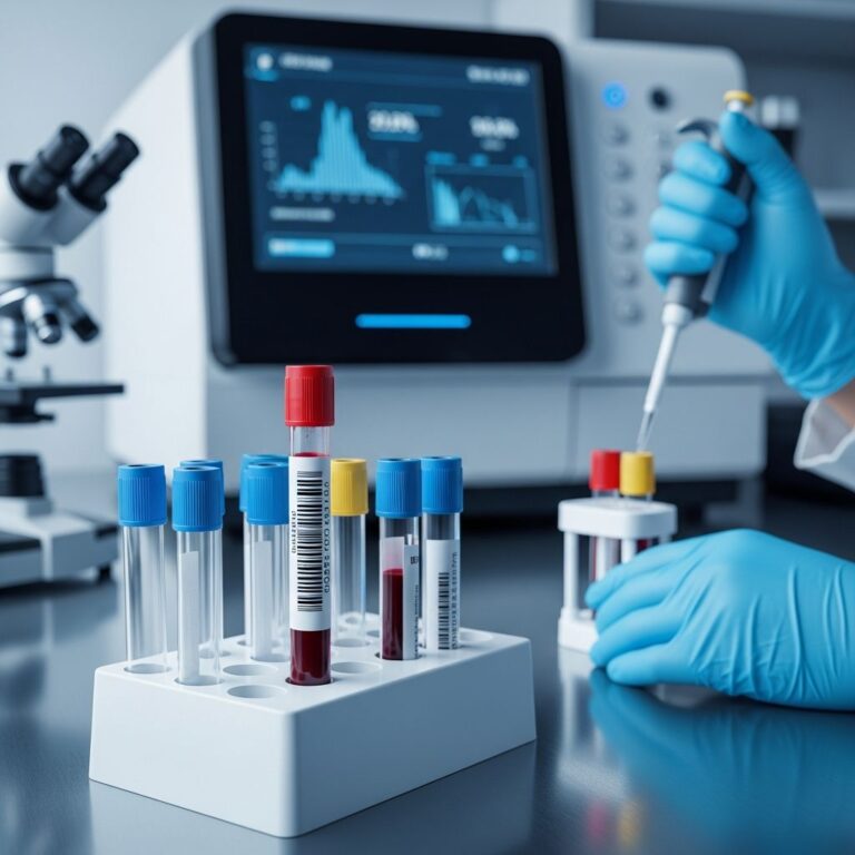
Histopathology after nail matrix biopsy
The biopsied tissue is fixed in formalin and processed for H&E staining, plus special stains (e.g., PAS for fungi, Melan-A for melanoma). Findings confirm diagnoses:
| Condition | Key Histopathologic Features |
|---|---|
| Psoriasis | Parakeratosis, Munro microabscesses, thinned suprapapillary plates. |
| Lichen planus | Band-like lymphocytic infiltrate, Civatte bodies, matrix hypergranulosis. |
| Melanoma | Atypical melanocytes in matrix epithelium, pagetoid spread. |
| Onychomatricoma | Filamentous digitations from matrix. |
Histopathology is gold standard, especially for onychomycosis (fungal invasion proof) or ruling out malignancy.
Frequently Asked Questions (FAQs)
Is nail matrix biopsy painful?
Local anesthesia makes the procedure painless; post-op discomfort is mild, managed with over-the-counter pain relievers.
Will my nail grow back normally?
Possibly, but permanent changes like ridges or splitting occur in some cases due to matrix scarring. Distal biopsies have better outcomes.
How long does recovery take?
Wound heals in 1-2 weeks; full nail regrowth: 3-6 months for fingers, longer for toes.
Can it diagnose fungal infections?
Matrix biopsy is for matrix issues; for onychomycosis, clip onycholyzed plate or proximal punch.
Is it done under general anesthesia?
No, local digital block suffices for outpatient setting.
Alternatives to nail matrix biopsy
Non-invasive options like dermoscopy, trichoscopy, or ultrasound guide but cannot replace histopathology for definitive diagnosis. Nail clipping suits distal issues; full avulsion for extensive disease.
In summary, nail matrix biopsy, though risky, is indispensable for matrix pathologies, balancing diagnostic yield against potential dystrophy.
References
- Nail Biopsy: A User’s Manual — Grover C, et al. Indian Dermatol Online J. 2018-01-15. https://pmc.ncbi.nlm.nih.gov/articles/PMC5803938/
- Nail Biopsy and Surgery — Comprehensive Dermatology Center. 2023. https://www.compdermcenter.com/nail-biopsy-and-surgery/
- Nail Biopsy — Perri Dermatology. 2010-08-08. https://perridermatology.com/dr-perris-blog/nail-biopsy/
- Nail Biopsy – StatPearls — Tschandl P. NCBI Bookshelf. 2023. https://www.ncbi.nlm.nih.gov/books/NBK567752/
- Playdough Surgery – Nail Matrix Biopsy — The Breakfasteur. YouTube. 2019. https://www.youtube.com/watch?v=pkF9Q308qVA
Similar Articles


Read full bio of medha deb