Undefined Necrobiosis Lipoidica Pathology: Histology Guide
Detailed histopathological analysis of necrobiosis lipoidica, a granulomatous skin disorder linked to diabetes and collagen degeneration.

Necrobiosis lipoidica (NL) is a chronic granulomatous dermatosis characterised by areas of necrobiosis (collagen degeneration) of the dermis that become surrounded by palisading granulomatous inflammation. It is strongly associated with diabetes mellitus, although up to 35% of patients with NL do not have diabetes.
What is necrobiosis lipoidica?
Necrobiosis lipoidica is an uncommon disorder of unknown aetiology in which the dermis shows degenerate collagen surrounded by palisading granulomas and chronic inflammation. Areas of fibrosis develop later. The epidermis is normal. Involvement of subcutaneous fat and blood vessels may occur.
The condition typically presents as yellow-brown plaques with telangiectasia on the shins of young adults with diabetes mellitus. However, lesions may occur at other sites and in the absence of diabetes.
Histology of necrobiosis lipoidica
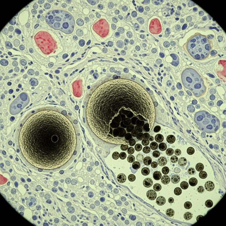
Scanning power view of necrobiosis lipoidica demonstrates a layered inflammatory process and alternating zones of necrobiosis involving the full thickness of the dermis (Figure 1). The changes tend to become more pronounced deeper in the dermis and may extend into the septal panniculus (Figures 2 and 3).
The areas of necrobiosis are poorly defined and run into each other with broad foci of inflammatory infiltrate intervening (Figure 4). This may form a stacked ‘lasagne’ type appearance. A variable histiocytic infiltrate with multinucleated giant cells surrounds these areas. The accompanying inflammatory infiltrate is predominantly lymphocytic with plasma cells and occasional eosinophils (Figure 5).
As lesions age an increasing degree of dermal fibrosis is seen.
Degenerate collagen – necrobiosis
The necrobiotic areas consist of eosinophilic homogeneous or slightly fibrillary collagen bundles with loss of nuclei and separation of collagen bundles by oedema and mucin. There is often horizontal layering of the necrobiotic collagen imparting a ‘layer cake’ appearance.
- Degenerate collagen bundles
- Loss of fibroblast nuclei
- Horizontal layering / ‘layer cake’ appearance
- Edema and mucin deposition
Granulomatous inflammation
Palisading histiocytes and multinucleate giant cells surround the areas of necrobiosis. Lymphocytes, plasma cells and eosinophils are present throughout the dermis.
- Palisading granulomas
- Multinucleated giant cells
- Mixed inflammatory cells (lymphocytes, plasma cells, eosinophils)
Fibrosis
In late lesions there is prominent sclerosis of the dermis with thick hyalinised collagen bundles arranged horizontally. Vessels show hyalinisation of walls.
Vascular changes
Dermal blood vessels show hyalinisation of walls, endothelial swelling and occasional luminal fibrin. Extravasated red blood cells may be seen.
Histopathology images
(Note: In a full HTML page, images would be embedded here showing scanning magnification, low power, necrobiosis zones, granulomatous inflammation, and fibrosis.)
- Figure 1: Scanning power view showing full thickness dermal involvement with layered necrobiosis.
- Figure 2: Degenerate collagen extending into subcutaneous fat.
- Figure 3: Layered ‘lasagne-like’ necrobiosis zones.
- Figure 4: Broad zones of necrobiosis with intervening inflammation.
- Figure 5: Palisading histiocytes, giant cells and mixed inflammatory infiltrate.
Differential diagnosis
The palisading granulomatous pattern in necrobiosis lipoidica must be distinguished from other necrobiotic granulomatous dermatitides.
| Feature | Necrobiosis lipoidica | Granuloma annulare | Rheumatoid nodule | Necrobiotic xanthogranuloma |
|---|---|---|---|---|
| Depth | Full dermis to subcutis | Superficial/mid dermis | Dermis/subcutis | Mid dermis/panniculus |
| Mucin | Little | Abundant | Absent | Small amounts |
| Normal dermis between foci | Absent | Present | Variable | Variable |
| Plasma cells | Common | Rare | Variable | Present |
| Cholesterol clefts | Rare | Absent | Absent | Common |
| Giant cells | Langerhans/foreign body | Langerhans | Absent | Toutons present |
Granuloma annulare
More superficial involvement with increased interstitial mucin that is Alcian blue positive. Normal dermis separates foci of necrobiosis. Fewer plasma cells.
Rheumatoid nodule
Central area of fibrinoid necrosis surrounded by palisading histiocytes. More subcutaneous involvement. Clinical correlation essential.
Necrobiotic xanthogranuloma
Touton’s giant cells and cholesterol clefts prominent. Associated with paraproteinaemia. Periorbital distribution.
Other differentials
- Sarcoidosis: ‘Naked’ granulomas without necrobiosis
- Foreign body reaction: Polariscopic material
- Infectious granulomas: Special stains for organisms
Clinical features
Understanding the pathology informs clinical recognition of necrobiosis lipoidica.
- Shiny yellow-brown plaques on pretibial skin
- Telangiectasia and atrophy
- Female predominance (3:1)
- Associated with diabetes (60-80% cases)
- Asymptomatic or tender; prone to ulceration
Who gets necrobiosis lipoidica?
NL most commonly affects young adults in their 30s, with a strong female predominance. Approximately 60% of patients have or will develop diabetes mellitus, often type 1.
Investigations
Diagnosis is confirmed by skin biopsy showing characteristic histopathology. Blood glucose and HbA1c should be checked in all patients.
Treatment of necrobiosis lipoidica
Treatment is often disappointing. Optimise diabetes control. First-line options include:
- Topical/intralesional corticosteroids
- Topical calcineurin inhibitors (tacrolimus)
- Anti-TNF agents (e.g., adalimumab)
Lesions may ulcerate and rarely undergo malignant transformation to squamous cell carcinoma.
Frequently Asked Questions
Q: What is the key histopathological feature of necrobiosis lipoidica?
A: Layered zones of necrobiosis involving the full thickness of the dermis with palisading granulomas, forming a characteristic ‘layer cake’ or ‘lasagne’ appearance.
Q: How does necrobiosis lipoidica differ from granuloma annulare on biopsy?
A: NL shows deep/full dermal involvement, little mucin, no normal dermis between foci, and common plasma cells. GA is more superficial with abundant mucin and normal intervening dermis.
Q: Is diabetes always present in necrobiosis lipoidica?
A: No, while strongly associated (60-80% cases), up to 35% of patients with NL do not have diabetes mellitus.
Q: What vascular changes are seen in necrobiosis lipoidica pathology?
A: Dermal vessels show hyalinisation of walls, endothelial swelling, and occasional fibrin thrombi.
Q: Can necrobiosis lipoidica undergo malignant transformation?
A: Yes, rarely squamous cell carcinoma may develop in long-standing ulcerated lesions.
This comprehensive review covers the essential pathological features of necrobiosis lipoidica, aiding accurate diagnosis and management. Early biopsy confirmation is crucial for distinguishing from mimics and guiding therapy.
References
- Necrobiosis lipoidica pathology image — DermNet NZ. 2023. https://dermnetnz.org/imagedetail/18304-necrobiosis-lipoidica-pathology
- Necrobiosis lipoidica — DermNet NZ. 2023. https://dermnetnz.org/topics/necrobiosis-lipoidica
- Necrobiosis lipoidica — Libre Pathology. 2024. https://librepathology.org/wiki/Necrobiosis_lipoidica
- Necrobiosis Lipoidica — Patient.info. 2023. https://patient.info/doctor/dermatology/necrobiosis-lipoidica-pro
- Necrobiotic xanthogranuloma pathology — DermNet NZ. 2023. https://dermnetnz.org/topics/necrobiotic-xanthogranuloma-pathology
- Necrobiosis Lipoidica — StatPearls, NCBI Bookshelf. 2023-10-01. https://www.ncbi.nlm.nih.gov/books/NBK459318/
Similar Articles


Read full bio of Sneha Tete