Necrobiosis Lipoidica: Key Insights On Symptoms And Treatment
Rare granulomatous skin disorder linked to diabetes, featuring shiny yellow-red plaques on shins with risk of ulceration.

What is necrobiosis lipoidica?
Necrobiosis lipoidica (NL) is a rare chronic granulomatous dermatitis characterised by collagen degeneration in the dermis, resulting in distinctive yellowish plaques with telangiectatic borders, most commonly on the pretibial areas of the lower legs. It affects women more frequently than men, with a peak onset between ages 30 and 40. Although strongly associated with diabetes mellitus, up to 35% of cases occur in non-diabetics, and blood glucose control does not influence disease progression.
The condition involves necrobiosis, a process of collagen degeneration, surrounded by granulomatous inflammation including palisading histiocytes, multinucleated giant cells, and lymphocytes. Horizontal layers of degenerated collagen extend from the superficial to deep dermis, accompanied by thickened vessel walls and perivascular inflammation. Fat deposition (lipoidica) contributes to the waxy appearance.
NL progresses slowly through stages: initial inflammatory plaques expand centrifugally, develop central atrophy with a ‘porcelain-like’ sheen, and may ulcerate following minor trauma. Lesions are often bilateral and symmetric on shins but can appear on thighs, arms, trunk, or face (rarely).
Who gets necrobiosis lipoidica?
NL has an incidence of approximately 0.3–1 per 1000 diabetics, comprising 1–2% of referrals to dermatology clinics specialising in diabetes-related skin conditions. Females are affected 3–4 times more often than males. About 60–80% of patients have or develop diabetes mellitus (type 1 or 2), with onset preceding diabetes diagnosis in 20% of cases. Familial predisposition exists, and paediatric cases are documented.
- Demographics: Predominantly adults 30–40 years; rare in children.
- Risk factors: Diabetes (11–65% association), autoimmune thyroid disease (25%), hypertension, hyperlipidaemia, obesity, metabolic syndrome.
- Non-diabetic cases: 20–40%, often with other autoimmune conditions like coeliac disease or sarcoidosis.
Comorbidities heighten vigilance: one study reported thyroid disease in 25% regardless of diabetes status, hypertension in 30%, and increased cardiovascular risk.
What causes necrobiosis lipoidica?
The aetiology remains idiopathic, but multifactorial hypotheses predominate. Diabetic microangiopathy is central: small vessel wall thickening, hyalinisation, and endothelial damage mirror retinal/kidney changes in diabetes. Basement membrane-like material accumulates similarly.
Immune dysregulation features prominently: defective neutrophils may hyperactivate macrophages, forming palisaded granulomas. Elevated tumour necrosis factor-alpha (TNF-α) in lesional skin and serum correlates with inflammation, akin to granuloma annulare.
Other contributors include:
- Trauma-induced koebnerisation (new lesions at injury sites).
- Genetic factors (familial clusters).
- Vascular occlusion from platelet aggregation or fibrinogen deposition.
No single trigger explains all cases; interplay of metabolic, vascular, and inflammatory pathways drives pathogenesis.
What are the clinical features of necrobiosis lipoidica?
Lesions begin as 2–5 mm red-brown papules or nodules on shins, expanding to 6–20 cm plaques with active erythematous borders and telangiectasias. Central areas atrophy, acquiring a yellow-waxy ‘glazed porcelain’ sheen, depressed 1–2 mm below surrounding skin. Hair loss, anhidrosis, and reduced sensation occur within plaques.
Symptoms vary: asymptomatic (majority), pruritic, painful, or paraesthetic. Ulceration affects 30% after minor trauma/pressure, healing with atrophic scarring. Rare sites include upper limbs, trunk, scalp, or periorbital (‘necrobiosis lipoidica diabeticorum et ulcerosa’). Giant NL exceeds 20 cm.
| Stage | Features |
|---|---|
| Inflammatory | Red papules/plaques, indurated violaceous border, telangiectasia. |
| Sclerosing/plaque | Yellow centre, waxy atrophy, telangiectasia prominent. | Ulcerative | Punched-out ulcers, haemosiderin pigmentation, scarring. |
Diagnosis
Clinical diagnosis suffices for typical pretibial porcelain-yellow plaques with telangiectasia in diabetic patients. Differential includes granuloma annulare (smaller, annular), erythema nodosum (painful nodules), sarcoidosis, or pretibial myxoedema.
Skin biopsy confirms: vertical necrobiotic zones of degenerated collagen/fibrin surrounded by palisaded histiocytes, giant cells, lymphoid infiltrate. Vessels show hyalinisation; mucin is scant. Direct immunofluorescence is negative.
Investigations screen comorbidities: fasting glucose/HbA1c, thyroid function (TSH, anti-TPO), lipids, full blood count. Doppler ultrasound assesses ulcers for vascular insufficiency.
What is the treatment for necrobiosis lipoidica?
Treatment is challenging, often disappointing; 30–50% remit spontaneously over years, but ulcers persist. Goals: halt progression, heal ulcers, prevent complications. No therapy alters established atrophy.
First-line: Potent/super-potent topical corticosteroids (e.g., clobetasol 0.05%) for early inflammatory lesions, applied 12–30g/week under occlusion. Monitor for atrophy/ulceration.
Intralesional: Triamcinolone acetonide (10–20 mg/mL) into active borders every 4–6 weeks; effective but risks ulceration.
Calcineurin inhibitors: Topical tacrolimus 0.1% or pimecrolimus bid; superior to steroids in some trials, safe for atrophic skin, excels in ulcers.
Advanced therapies:
- Antimalarials: Hydroxychloroquine 200–400 mg/day; 50–70% response in small series.
- Anti-TNF: Adalimumab/infliximab; meta-analysis shows 70% complete regression.
- Phototherapy: PUVA/NB-UVB (50% response); photodynamic therapy for refractory cases.
- Others: Methotrexate, ciclosporin, dapsone, mycophenolate; hyperbaric oxygen for ulcers.
Ulcer management: multilayer compression, pentoxifylline/aspirin/dipyridamole, debridement. Surgical excision/grafting for non-healing ulcers; laser (pulsed dye) improves cosmesis.
Adjuncts: smoking cessation, trauma avoidance, weight loss, diabetes control (despite limited impact).
Complications
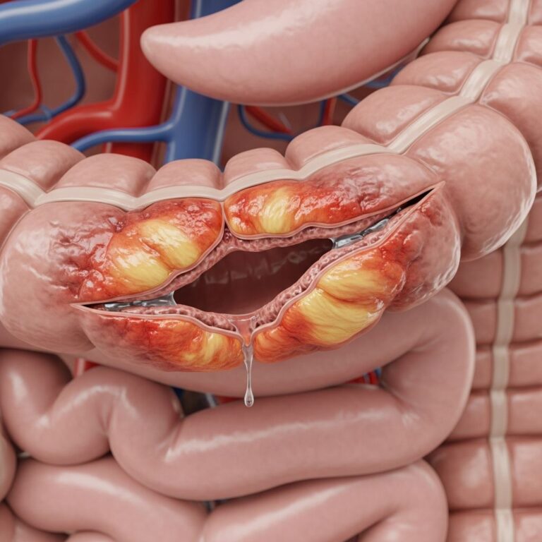
Ulceration (25–35%) leads to infection, scarring, lymphoedema. Rare squamous cell carcinoma (1–2%) in chronic ulcers requires biopsy of non-healing lesions >3 months.
Prevention
No proven prevention; protect legs from trauma, optimise diabetes/metabolic control, screen high-risk patients.
Further reading and references
Consult dermatology for biopsy/trials. Patient education on leg care essential.
Frequently Asked Questions
Is necrobiosis lipoidica always associated with diabetes?
No, 20–40% occur in non-diabetics, though diabetes increases risk 10–40-fold.
Does treating diabetes cure NL?
No, glycaemic control does not improve lesions.
Can NL lesions become cancerous?
Rarely; squamous cell carcinoma arises in <2% of chronic ulcers—biopsy suspicious changes.
What is the best treatment for ulcers?
Multimodal: compression, tacrolimus, hyperbaric oxygen; surgery if refractory.
Will NL go away on its own?
30–50% remit spontaneously over 5–10 years, but recurrence common.
References
- Necrobiosis Lipoidica – MD Searchlight — MD Searchlight. 2023. https://mdsearchlight.com/skin-problems-and-treatments/necrobiosis-lipoidica/
- Necrobiosis lipoidica: Definition, causes, risk factors, and treatment — Medical News Today. 2023-10-12. https://www.medicalnewstoday.com/articles/necrobiosis-lipoidica
- Necrobiosis lipoidica diabeticorum – UCSF Health — UCSF Health. 2024. https://www.ucsfhealth.org/medical-tests/necrobiosis-lipoidica-diabeticorum
- Necrobiosis lipoidica — RACGP. 2014-03-01. https://www.racgp.org.au/afp/2014/march/necrobiosis-lipoidica
- Necrobiosis lipoidica – DermNet — DermNet NZ. 2025. https://dermnetnz.org/topics/necrobiosis-lipoidica
- necrobiosis lipoidica — National Organization for Rare Disorders. 2024. https://rarediseases.org/mondo-disease/necrobiosis-lipoidica/
- Necrobiosis Lipoidica – StatPearls — NCBI Bookshelf. 2023-07-17. https://www.ncbi.nlm.nih.gov/books/NBK459318/
Similar Articles


Read full bio of medha deb