Necrolytic Migratory Erythema: Expert Guide To Diagnosis & Treatment
Understanding the characteristic rash of glucagonoma syndrome: causes, symptoms, diagnosis, and effective treatments.

Necrolytic migratory erythema (NME) is a distinctive cutaneous manifestation primarily associated with glucagonoma syndrome, caused by a rare, slow-growing malignant tumour in the alpha cells of the pancreas.
What is necrolytic migratory erythema?
Necrolytic migratory erythema represents the hallmark dermatological feature of glucagonoma syndrome, occurring in approximately 90% of affected individuals. This syndrome arises from excessive glucagon secretion by the pancreatic alpha-cell tumour, known as glucagonoma, which predominantly impacts adults over 50 years of age. The tumour’s hypersecretion disrupts metabolic homeostasis, leading to characteristic skin eruptions alongside systemic effects such as diabetes mellitus and profound weight loss.
While classically linked to glucagonoma, NME-like rashes can emerge in pseudoglucagonoma syndrome, stemming from conditions like malabsorption, liver cirrhosis, or inflammatory bowel disease, where glucagon levels remain normal. The precise pathogenesis remains elusive but is attributed to relative deficiencies in zinc, essential amino acids, and fatty acids, compounded by hypoaminoacidaemia and hypoalbuminemia, which impair nutrient transport. Hyperglucagonaemia may exacerbate cutaneous inflammation, particularly in friction-prone areas, while elevating blood glucose and catabolizing proteins and fats.
Clinical features
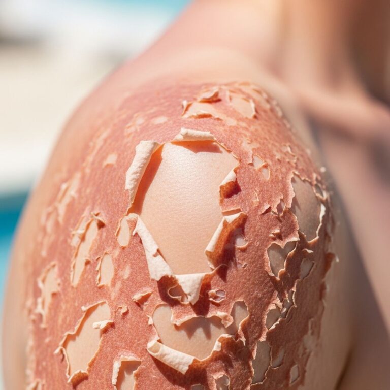
NME manifests as polymorphic, migratory lesions that evolve through distinct phases: initial annular erythema, central vesiculation, erosion, crusting, and peripheral extension with healing at the core. Lesions predominantly localize to intertriginous zones including the groin, perineum, buttocks, lower abdomen, and thighs, with occasional involvement of distal extremities, face, or trunk.
The rash exhibits a cyclical pattern, waxing and waning over 7–14 days, often triggered or worsened by trauma. Affected areas display erythematous plaques with scaling, blistering, and secondary crusting, accompanied by intense pruritus or burning pain. Healing leaves post-inflammatory hyperpigmentation. Mucosal involvement is common, featuring a painful, atrophic glossitis, angular cheilitis, and nail dystrophy with Beau’s lines.
- Erythematous, annular plaques with advancing borders
- Central blistering, erosion, and crusting
- Pruritus and pain, especially in flexures
- Mucocutaneous changes: glossitis, cheilitis, nail ridging
- Cyclical migration every 10 days on average
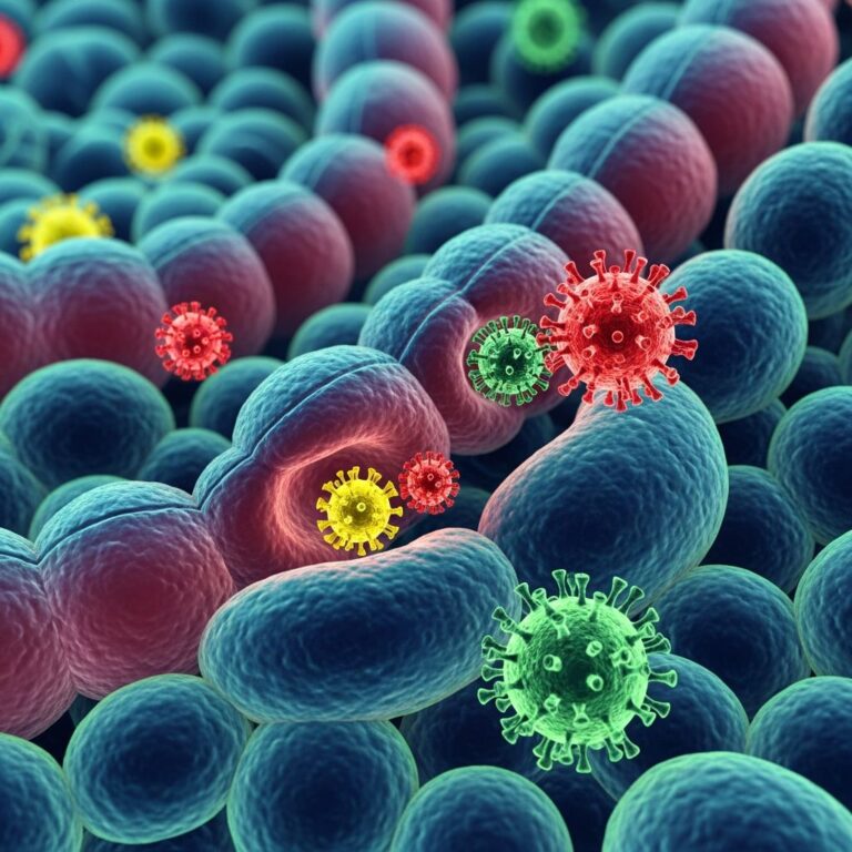
- Secondary infection with Staphylococcus aureus or Candida, forming pustules
Systemic glucagonoma syndrome features include new-onset diabetes (30–50% at diagnosis), cachexia (70%), anaemia, diarrhoea, deep vein thrombosis, and neuropsychiatric symptoms.
Associated conditions
Beyond glucagonoma, NME resembles eruptions in:
- Zinc deficiency (acrodermatitis enteropathica)
- Essential fatty acid or amino acid malnutrition
- Liver disease or enteric malabsorption (pseudoglucagonoma)
Complications
NME itself poses minimal direct complications, but glucagonoma syndrome engenders:
- Metabolic derangements: hypoaminoacidaemia, hypoalbuminemia, hyperglycaemia
- Cachexia and malnutrition
- Diabetes mellitus
- Thromboembolic events
- Secondary infections of lesions
Untreated, 50% mortality occurs within 5 years post-diagnosis due to tumour metastasis.
Differential diagnosis
NME’s migratory, annular morphology prompts consideration of:
| Condition | Key Distinguishing Features |
|---|---|
| Acrokeratosis paraneoplastica (Bazex syndrome) | Psoriasis-like acral keratoderma; associated with upper aerodigestive carcinomas |
| Subcorneal pustular dermatosis | Flaccid pustules in annular/circinate patterns; neutrophilic on biopsy |
| Pemphigus foliaceus | Superficial acantholysis; Nikolsky-positive |
| Psoriasis | Stable plaques without migration; Auspitz sign |
| Tinea corporis | Responds to antifungals; KOH-positive hyphae |
| Cellulitis | Acute fever, leukocytosis; bacterial aetiology |
| Erythrokeratoderma | Generalized hyperkeratosis from birth |
Investigations
Suspected NME mandates urgent endocrinological and oncological evaluation. Baseline tests reveal:
- Hyperglucagonaemia (>1000 pg/mL, often >10,000 pg/mL)
- Glucose intolerance/diabetes
- Hypoaminoacidaemia (all non-essential amino acids low)
- Anaemia, hypoalbuminemia
- Zinc deficiency
Skin biopsy demonstrates necrolytic epidermal separation: pale upper epidermis, necrotic keratinocytes, and diminished granular layer. Imaging localizes the pancreatic tail tumour:
- CT/MRI abdomen (preferred)
- Endoscopic ultrasound
- 68Ga-DOTATATE PET-CT for somatostatin receptor expression
Management
Treatment targets the underlying glucagonoma, with rapid rash amelioration post-resection in localized disease.
Surgical
Curative pancreatectomy for non-metastatic tumours (<2 cm, no lymph nodes).
Medical
For metastatic disease (60–80% at diagnosis):
- Somatostatin analogues: Octreotide LAR (30 mg IM monthly) or lanreotide; inhibit glucagon, resolve NME in 70–90% within weeks
- Nutritional support: IV amino acids, zinc (up to 220 mg/day), essential fatty acids; dramatic rash improvement
- Chemotherapy: Streptozotocin + 5-FU; dacarbazine; limited efficacy
- Targeted therapies: Everolimus, sunitinib for progressive disease
- PRRT: Peptide receptor radionuclide therapy
- Embolization/RFA for liver metastases
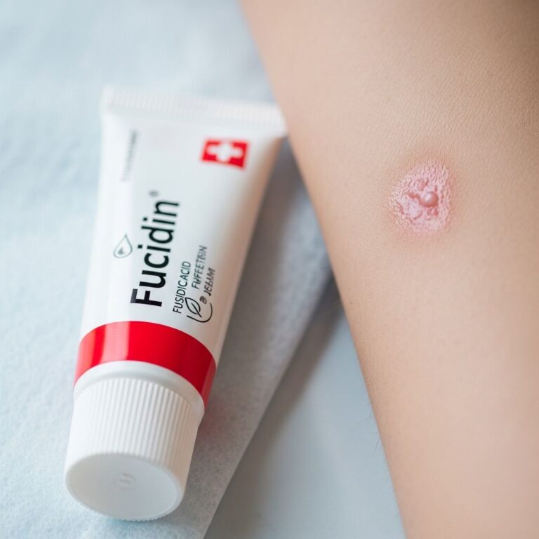
Symptomatic relief: topical corticosteroids, calcipotriol, oral tetracyclines for pustular variants.
Prognosis
Excellent post-resection (5-year survival >80%); median survival 3–5 years with metastases, improved by somatostatin analogues. NME recurs with tumour progression but resolves promptly with effective therapy.
Frequently Asked Questions
What causes necrolytic migratory erythema?
NME is primarily caused by glucagonoma, a rare pancreatic alpha-cell tumour secreting excess glucagon. Pseudoglucagonoma forms occur with malnutrition or liver disease.
How is NME diagnosed?
Diagnosis combines clinical morphology, hyperglucagonaemia, characteristic biopsy (necrolytic changes), and pancreatic imaging (CT/MRI).
Does NME rash itch?
Yes, lesions cause significant pruritus and pain, particularly during blistering/erosive phases.
Can NME be cured?
Curative surgery for localized glucagonoma resolves NME; metastatic cases require lifelong somatostatin therapy.
What is the prognosis for glucagonoma?
Localized: excellent; metastatic: 3–5 years median survival with modern therapies.
References
- Necrolytic Migratory Erythema — MD Searchlight. 2023. https://mdsearchlight.com/skin-problems-and-treatments/necrolytic-migratory-erythema/
- Necrolytic migratory erythema (glucagonoma syndrome) — DermNet NZ. 2004 (updated). https://dermnetnz.org/topics/necrolytic-migratory-erythema
- Necrolytic migratory erythema — Primary Care Dermatology Society (PCDS). 2021-07-26. https://www.pcds.org.uk/clinical-guidance/necrolytic-migratory-erythema
- Glucagonoma: What It Is, Causes, Symptoms & Treatment — Cleveland Clinic. 2023. https://my.clevelandclinic.org/health/diseases/25246-glucagonoma
- Necrolytic Migratory Erythema — StatPearls [Internet]. NCBI Bookshelf. 2023. https://www.ncbi.nlm.nih.gov/books/NBK532872/
Similar Articles


Read full bio of medha deb