Necrotising Infundibular Crystalline Folliculitis Pathology
Understanding the pathology and histopathological features of NICF in dermatology.

Necrotising infundibular crystalline folliculitis (NICF) is a distinctive and rare folliculocentric disorder characterized by specific clinical and histopathological features that differentiate it from other follicular conditions. This condition presents with sharply demarcated waxy papules that typically appear in seborrheic areas of the skin. The disorder is defined by the accumulation of unusual material within hair follicles, which is composed of elements derived from Malassezia yeasts, bacteria, and sebaceous lipids. Understanding the pathology of NICF requires examination of both its clinical presentation and the underlying histopathological changes that occur within affected follicles.
Clinical Presentation of NICF
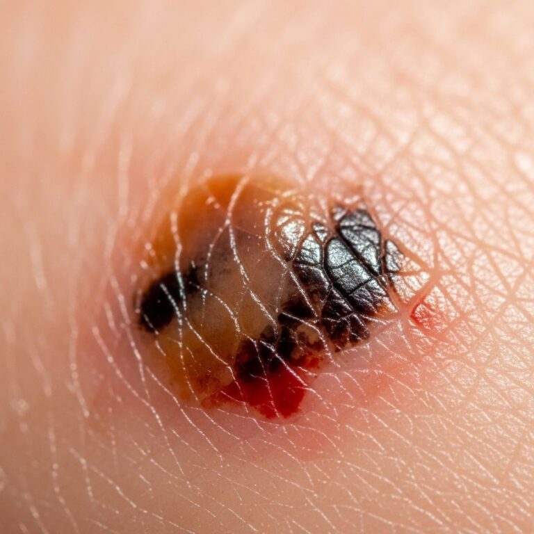
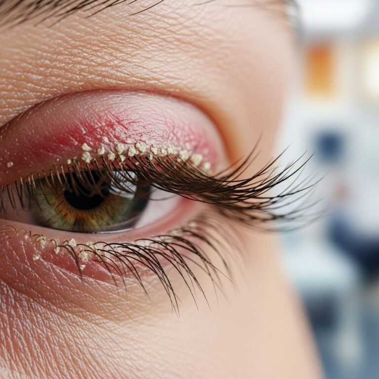
The clinical appearance of necrotising infundibular crystalline folliculitis is highly distinctive and recognizable to trained dermatologists. Patients typically present with the sudden appearance of numerous pale waxy filamentous follicular plugs that are matted together. These lesions are characteristically localized to seborrheic areas, particularly the forehead and upper back regions of the body. The papules are sharply demarcated and have a distinctive waxy appearance that helps clinicians identify the condition during examination.
The clinical presentation is relatively uniform across affected individuals, with eruptive yellow waxy umbilicated folliculocentric plugs appearing as the primary manifestation. The sudden onset of numerous lesions simultaneously is a hallmark feature of this disorder, distinguishing it from other chronic follicular conditions that develop gradually over time. The lesions themselves are follicular-based and appear as pale, waxy material protruding from the skin surface.
Histopathological Features and Findings
The histopathology of NICF reveals distinctive microscopic features that are essential for accurate diagnosis. When sections are examined under the microscope, filamentous deposits become evident, and these are enclosed by parakeratotic columns within a partly necrotic follicle ostium. The follicular opening shows characteristic changes with necrotic material present alongside the abnormal deposits.
At higher magnification, crystalline urate-like structures become visible within the filamentous material present inside the follicles. These crystalline formations are a defining feature of the condition and contribute to its name. The material demonstrates birefringence when examined under polarized light, which is a key diagnostic characteristic that helps differentiate NICF from other follicular disorders.
Composition of Follicular Material
The material found within affected follicles in NICF cases consists of pale crystalline filaments embedded in an amorphous sebum-rich substance. This composition is remarkable because, despite the presence of abundant sebaceous material, recognizable structured sebaceous glands are typically absent in the affected areas. The histopathology is dominated by sebum production, yet the normal glandular architecture is not present.
Notably, only the superficial infundibular ostia (openings) remain visible, and the distended cavity beneath is devoid of follicular or sebaceous gland remnants. This represents a fundamental departure from the normal follicular structure, where sebaceous glands should be present and functional. The absence of these normal structures while sebaceous material accumulates creates the unique histopathological picture characteristic of NICF.
Microscopic Changes in the Follicular Wall
The follicular epithelium in NICF shows specific changes that are important for diagnosis. Parakeratotic epithelial lips are recognizable at the follicle opening, and the remaining histopathological features that typically define sebaceous follicles are usually no longer apparent. This loss of normal follicular architecture is a distinctive feature that helps pathologists identify NICF under the microscope.
The partly necrotic nature of the follicle ostium indicates that tissue death and destruction are occurring within the follicular structure itself. This necrotic component is integral to the condition’s name and reflects the destructive process affecting the follicle. The rupture of follicular epithelium and neutrophilic inflammation also contribute to the histopathological picture in some cases.
Etiology and Pathogenesis
The pathogenesis of NICF remains incompletely understood, though several factors have been identified as contributing to its development. The follicular accumulation of abnormal material is thought to be derived from multiple sources including Malassezia yeasts, bacteria, and sebaceous lipids. These components interact within the follicular environment to create the distinctive material seen in NICF.
NICF has been increasingly recognized as a paradoxical side effect of certain antitumor inhibitors, particularly those targeting epidermal growth factor receptor (EGFR), vascular endothelial growth factor (VEGF), and programmed death-1 (PD-1) pathways. The emergence of NICF in patients receiving these targeted therapies represents an expression of altered molecular pathways that underpin the condition’s development. These medications, while treating malignancy, can disrupt the normal homeostatic mechanisms that maintain follicular health.
Role of Infundibular Stem Cells
A critical aspect of NICF pathogenesis involves committed infundibular stem cells. Infundibulogenesis, the formation of the follicular infundibulum, depends on the activation of these specialized stem cells. The infundibulum normally has an epidermoid lining that is integrated with the interfollicular epidermis, maintaining a consistent pattern of differentiation with surrounding skin.
When unscheduled rapid amplification occurs in the infundibular committed stem cell pathway, there is potential for failure to form differentiated sebaceous glands properly. The resulting aberrant sebum-producing infundibular stem cells may undergo clonal expansion and subsequently disintegrate as a result of holocrine sebum production, leading to total loss of infundibular integrity. This represents an abortive sebaceous folliculogenesis process that distinguishes NICF from normal follicular development.
Relationship to Crystalline Formation
The terminal production of polarizable crystalline concretions represents a significant aspect of NICF pathology. These crystalline formations result from the consumption and loss of normal follicular structures through the holocrine sebum production process. The crystalline structures visible under polarized light are composed of urate-like material that accumulates as the follicle undergoes its pathological changes.
The presence of yeast organisms, particularly Malassezia species, within the sebaceous material may play a role in crystalline formation. PAS staining can demonstrate pityrosporum species within the follicular material, confirming the presence of fungal elements in some cases. These microorganisms may contribute to the chemical composition or precipitation of crystalline material within the follicle.
Diagnostic Considerations and Differential Diagnosis
Accurate diagnosis of NICF requires correlation of clinical presentation with histopathological findings. The distinctive clinical appearance of pale waxy follicular plugs combined with the characteristic microscopic features of crystalline deposits and parakeratotic columns allows for definitive identification of the condition. Pathologists examining tissue samples must recognize the combination of features that define NICF.
Differentiation from Other Conditions
Several other conditions must be considered in the differential diagnosis of NICF. Gout represents an important differential consideration because the distinctive filamentous material in NICF resembles the crystalline deposits seen in cutaneous gout. However, the follicular localization and additional histopathological features of NICF help distinguish it from systemic gout manifesting in the skin.
Other follicular and keratotic disorders must also be considered when evaluating patients with follicular plugs. The waxy appearance and specific localization to seborrheic areas help differentiate NICF from keratosis pilaris, acanthosis nigricans, and other common follicular conditions. The histopathological examination revealing crystalline deposits within a necrotic follicle ostium is essential for definitive differentiation.
Incidental Crystalline Findings
Interestingly, crystalline structured sebum-rich material similar to that seen in eruptive NICF has been observed as an incidental feature in intact sebaceous follicles. This finding typically occurs within cystically dilated, fully differentiated sebaceous follicles or as limited crystalline plugs at the orifice of intact sebaceous follicles. However, these incidental findings lack the necrotizing aspects characteristic of eruptive NICF, showing preservation of both infundibular and mature sebaceous gland differentiation.
This distinction is clinically important because it suggests that crystalline material can form in normal follicular settings without the pathological consequences seen in true NICF. The development of actual necrotizing folliculitis with loss of both infundibular and sebaceous gland differentiation represents a more severe process than simple crystalline accumulation.
Resolution and Clinical Course
The resolution of NICF is enhanced by the presence of open expanded infundibular orifices that provide a passage for the elimination of sebaceous concretions. These open follicular openings allow material to drain from the affected follicles, facilitating recovery. This drainage mechanism is an important component of the natural healing process in NICF.
The rare phenomenon of NICF may represent an aberrant pathway with disrupted sebaceous follicular histogenesis due to a loss of homeostatic control. The condition appears to require a series of synchronized events inherent in the committed infundibular stem cell pathway, linked to the healing phase of folliculitis, along with temporary activating triggers such as a mix of organisms in sebum-rich follicles. The presence and proper function of regulatory T cells in maintaining homeostatic integrity of sebaceous follicles and establishing immune tolerance to skin commensal microbes may also play important roles in NICF development and resolution.
Special Considerations in Drug-Induced NICF
Destruction and repair of the follicular infundibulum itself appears to be integral to activating the committed infundibular stem cell pathway leading to NICF. The unscheduled modulation of the infundibular homeostatic sebaceous repair axis by EGFR inhibitors, VEGF inhibitors, and PD-1 inhibitors may lead to an aberrant outcome with metaplasia of infundibular keratinocytes to sebocytes. When sebaceous gland differentiation fails to occur properly, these metaplastic infundibular sebocyte cells lead to consumption and loss of the infundibulum through holocrine sebum production.
Both VEGF and EGFR inhibitors have pustular and necrotizing folliculitis as common side effects, making recognition of NICF in this patient population particularly important. Clinicians prescribing these agents should monitor patients for the development of unusual follicular lesions that might represent drug-induced NICF.
Frequently Asked Questions
What is the primary clinical appearance of NICF?
NICF presents clinically with sharply demarcated waxy papules that appear suddenly in seborrheic areas, particularly the forehead and upper back. These lesions are pale, waxy follicular plugs that are often matted together and create a distinctive clinical appearance.
What are the key histopathological findings in NICF?
The key histopathological findings include filamentous deposits enclosed by parakeratotic columns within a partly necrotic follicle ostium. Crystalline urate-like structures are visible within the filamentous material, and the material is birefringent under polarized light. The normal sebaceous glands are typically absent despite abundant sebaceous material.
What causes NICF?
NICF is caused by follicular accumulation of material derived from Malassezia yeasts, bacteria, and sebaceous lipids. The condition has been linked to antitumor inhibitors targeting EGFR, VEGF, and PD-1 pathways. It appears to involve disruption of normal infundibular stem cell homeostasis and sebaceous follicle formation.
How is NICF differentiated from gout?
While the distinctive filamentous material in NICF resembles gout crystals, NICF is localized to hair follicles with characteristic parakeratotic columns around a necrotic follicle ostium. Gout lacks this follicular localization and histopathological pattern, making differentiation possible through clinical presentation and histopathological examination.
Is NICF related to medication use?
Yes, NICF has been increasingly recognized as a paradoxical side effect of antitumor inhibitors, particularly those targeting EGFR, VEGF, and PD-1 pathways. Both VEGF and EGFR inhibitors have pustular and necrotizing folliculitis as common side effects.
What special stains help diagnose NICF?
PAS staining can demonstrate Pityrosporum (Malassezia) species within the follicular material. Polarized light examination reveals the birefringence of the crystalline material, which is a key diagnostic feature of NICF.
References
- Eruptive Necrotizing Infundibular Crystalline Folliculitis — National Center for Biotechnology Information (NIH). 2021. https://pmc.ncbi.nlm.nih.gov/articles/PMC8601669/
- Necrotising infundibular crystalline folliculitis pathology — DermNet NZ. 2013. https://dermnetnz.org/topics/necrotising-infundibular-crystalline-folliculitis-pathology
- Necrotizing infundibular crystalline folliculitis: a clinicopathological study — PubMed. National Library of Medicine. https://pubmed.ncbi.nlm.nih.gov/22015152/
- Recurrent inverse necrotizing infundibular crystalline folliculitis — Journal of Cutaneous Pathology. Wiley Online Library. https://onlinelibrary.wiley.com/doi/abs/10.1111/cup.14617
- Necrotizing infundibular crystalline folliculitis — University of Zurich Research Archive. https://www.zora.uzh.ch/id/eprint/68907/
Similar Articles


Read full bio of medha deb