Nipple Papillomatosis: Causes, Diagnosis, and Treatment
Understanding benign nipple growths: clinical features, diagnosis, and management options.

Nipple Papillomatosis: A Comprehensive Clinical Overview
Nipple papillomatosis is a benign dermatological condition characterized by the growth of numerous papillomas arising from the lactiferous duct epithelium of the nipple. This relatively uncommon condition, also known as florid papillomatosis of the nipple, nipple adenoma, and erosive adenomatosis, presents as a distinct clinical entity that requires proper diagnosis and management. Understanding this condition is essential for both healthcare providers and patients to ensure appropriate treatment and peace of mind regarding the benign nature of the lesion.
Introduction and Classification
Nipple papillomatosis represents a specific benign proliferative disorder of the nipple epithelium. The condition is distinct from other breast papillomas, as it involves multiple papillomatous growths concentrated on the nipple surface rather than within the milk ducts themselves. This distinction is important for accurate diagnosis and appropriate management planning. The terminology surrounding this condition reflects its various presentations and has evolved over time, with multiple synonymous terms used in medical literature.
Demographics and Epidemiology
Nipple papillomatosis is classified as a rare condition, though the exact prevalence remains uncertain. The condition predominantly affects women, with peak incidence occurring in women aged 35 to 55 years. However, it is important to note that nipple papillomatosis can present at any age and occasionally affects men, indicating that age-related generalizations should not exclude individuals from diagnostic consideration. The demographic pattern suggests a potential hormonal or age-related predisposition, though the exact etiopathogenic mechanisms remain incompletely understood.
Clinical Features and Presentation
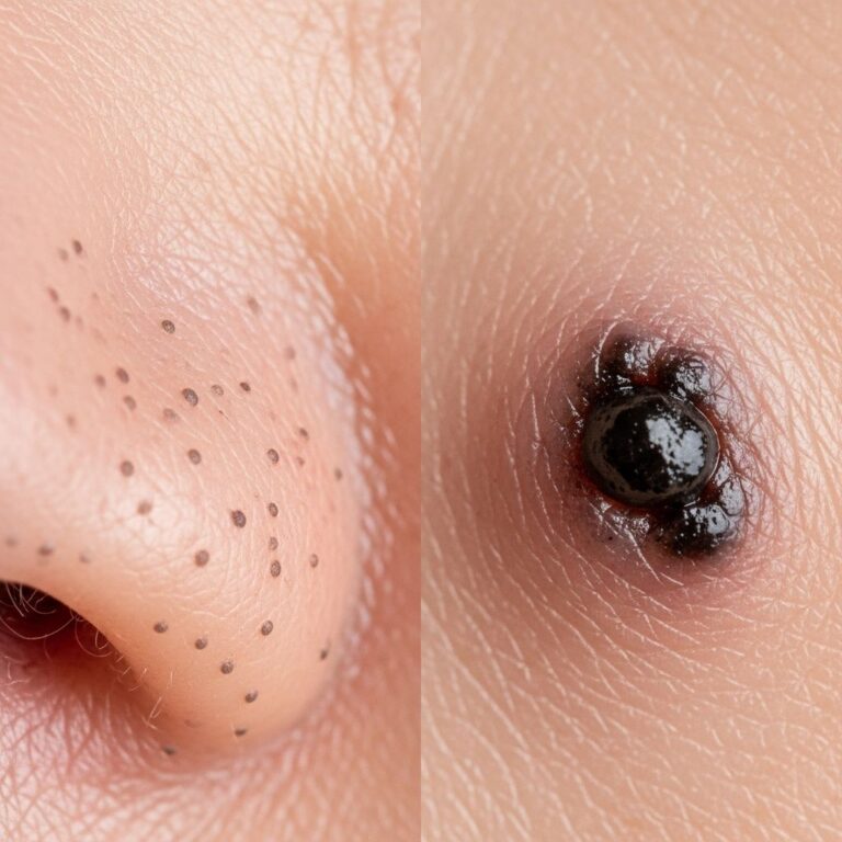
Nipple papillomatosis presents with characteristic clinical manifestations that may be noticed by patients or discovered during routine breast examination. The primary presentation typically includes:
- A palpable mass or swelling on the surface of the nipple
- Multiple small papillary projections creating a textured or cobblestone appearance
- Visible papillomatous growths on the nipple surface
- Potential nipple discharge, which may be clear, serous, or occasionally blood-tinged
- Mild nipple irritation or discomfort in some cases
- Incidental discovery during routine breast self-examination or clinical examination
The lesion may be asymptomatic in many cases, discovered only during a routine physical examination by a healthcare provider. The appearance can vary from subtle surface irregularities to more pronounced papillomatous changes. Some patients report noticing the condition because of changes in nipple appearance or texture, while others may be entirely unaware of its presence until professional evaluation.
Pathophysiology and Histopathological Features
The histopathological examination of nipple papillomatosis reveals distinctive architectural features that confirm diagnosis. Microscopically, the condition demonstrates proliferating ductal structures extending into the breast stroma, lined by a double layer of epithelium characteristic of normal ductal tissue. Additional histological features include the presence of keratin-filled cysts and tiny apical cell snouts, which are architectural elements that aid in distinguishing this condition from other nipple pathologies.
The epithelial proliferation in nipple papillomatosis represents a benign process without atypia or dysplasia in most cases. The double-layer epithelial lining maintains the structural integrity typical of normal ductal epithelium, differentiating it from malignant processes that often show disrupted epithelial architecture and loss of normal layering patterns.
Associated Variants and Differential Considerations
An important histological variant of nipple papillomatosis is syringomatous adenoma, which presents with distinctive pathological features. This variant is distinguished on histology by the absence of intraductal epithelial hyperplasia and the presence of oval, elongated ducts. Syringomatous adenoma tends to display more infiltrative and locally invasive characteristics compared to typical nipple papillomatosis, potentially requiring different management approaches. Recognition of this variant during histopathological examination helps guide appropriate clinical management decisions.
Diagnostic Evaluation and Imaging
Accurate diagnosis of nipple papillomatosis requires a systematic approach combining clinical examination, imaging studies, and histopathological confirmation. The diagnostic process typically includes:
- Detailed clinical history and physical examination of the affected nipple
- Assessment of nipple appearance, texture, and any associated discharge
- Breast imaging studies to evaluate for underlying lesions
- Histopathological confirmation through skin biopsy when diagnosis requires confirmation
Imaging studies such as breast X-ray, mammography, or ultrasound examination may be performed to exclude an underlying tumour or other significant breast pathology. Ultrasound is particularly useful in evaluating the extent of nipple involvement and assessing the deeper breast tissue to ensure no associated intraductal pathology. Mammography may be indicated in women above 40 years or those with additional breast cancer risk factors, as it provides a comprehensive assessment of the entire breast tissue and helps exclude occult malignancy.
A skin biopsy may be performed to confirm the diagnosis of nipple papillomatosis when clinical presentation is unclear or when histological confirmation is necessary. The biopsy specimen should be adequately sized to capture the full thickness of the lesion and surrounding normal tissue, allowing pathologists to make accurate diagnostic assessment and exclude malignant transformation.
Differential Diagnosis
Several conditions may present similarly to nipple papillomatosis and must be considered during diagnostic evaluation to ensure accurate diagnosis and appropriate management. The differential diagnosis for nipple papillomatosis includes:
- Intraductal papilloma—solitary or multiple growths within breast ducts that may present with nipple discharge
- Paget’s disease of the nipple—a malignant condition presenting with nipple changes and requiring urgent intervention
- Papillary carcinoma—a malignant breast lesion requiring different management
- Fibrocystic changes—benign breast changes that may appear similar clinically
- Nipple eczema or dermatitis—inflammatory conditions affecting the nipple
- Accessory breast tissue—ectopic breast tissue presenting as nipple abnormality
Distinguishing nipple papillomatosis from these conditions requires careful clinical assessment combined with imaging and histopathological evaluation. Paget’s disease of the nipple represents the most critical differential diagnosis to exclude, as this condition represents malignancy and requires aggressive intervention. Clinical features such as erosion, erythema, crusting, and associated ductal carcinoma in situ on biopsy help differentiate Paget’s disease from benign nipple papillomatosis.
Cancer Risk and Prognostic Considerations
Nipple papillomatosis is fundamentally a harmless, benign condition without malignant potential in itself. However, as with any fibrocystic change within breast tissue, there are reported associations with a slightly increased risk of breast cancer when compared with the general population. This increased risk, though notable from a statistical perspective, remains very low in absolute terms and should not cause undue concern in patients with this diagnosis.
The presence of nipple papillomatosis does not necessitate aggressive treatment purely based on cancer risk. Instead, management decisions should be individualized based on symptomatology, cosmetic concerns, and patient preferences. Regular breast health surveillance remains appropriate for all patients, particularly those with additional breast cancer risk factors.
Treatment Approaches and Management Strategies
Management of nipple papillomatosis should be individualized based on patient presentation, symptomatology, and preferences. Where practical, nipple papillomatosis is treated by complete excision of the lesion, which provides both therapeutic benefit and histopathological confirmation of diagnosis.
Surgical Treatment
Complete surgical excision represents the definitive treatment option for symptomatic nipple papillomatosis. This is typically a minor procedure usually performed under local anesthesia, making it accessible and well-tolerated by patients. The surgical approach involves careful removal of the entire affected area of the nipple, ensuring complete elimination of papillomatous tissue to prevent recurrence.
Surgical excision offers several advantages beyond therapeutic benefit. The removed tissue can be sent for histopathological examination, confirming diagnosis and definitively excluding malignancy. Complete excision also provides aesthetic improvement for patients concerned about nipple appearance and eliminates any associated symptoms such as irritation or discharge.
Conservative Management
Not all patients with nipple papillomatosis require immediate surgical intervention. If the entire affected area is not removed during initial treatment, or if patients opt for conservative management, the affected area should be reviewed from time to time, especially if the appearance changes. Regular clinical surveillance ensures that any changes in the lesion’s characteristics are detected promptly and appropriately managed.
Conservative observation may be appropriate for asymptomatic patients with diagnosis confirmed on biopsy who have minimal cosmetic concerns. Regular follow-up examination allows detection of any changes requiring intervention, while avoiding unnecessary surgery in patients who are comfortable with their diagnosis and presentation.
Post-Treatment Considerations
Following surgical excision, patients should expect normal wound healing with minimal scarring. The small surgical site typically heals well with appropriate wound care. Patients should follow post-operative instructions regarding wound care, activity restrictions, and follow-up appointments as directed by their surgeon.
Any persistent or recurrent symptoms should be reported to the healthcare provider for evaluation. Although recurrence is uncommon with complete excision, vigilance for new lesions or changes in the contralateral nipple remains important given the potential for bilateral involvement in some patients.
Frequently Asked Questions
Q: Is nipple papillomatosis cancer?
A: No, nipple papillomatosis is a benign, non-cancerous condition. While there is a slightly increased risk of breast cancer associated with fibrocystic changes in general, nipple papillomatosis itself does not transform into cancer.
Q: Who is most likely to develop nipple papillomatosis?
A: Nipple papillomatosis most commonly affects women aged 35 to 55 years, though it can occur at any age and occasionally affects men. The exact cause remains unknown.
Q: What are the main symptoms of nipple papillomatosis?
A: The primary symptom is a palpable mass or swelling on the nipple surface. Some patients experience nipple discharge or mild irritation, while others discover the condition incidentally during routine examination.
Q: How is nipple papillomatosis diagnosed?
A: Diagnosis involves clinical examination, imaging studies such as ultrasound or mammography, and confirmation through skin biopsy. Histopathological examination revealing proliferating ductal structures with double-layer epithelium confirms the diagnosis.
Q: Is treatment necessary for nipple papillomatosis?
A: Treatment is not always necessary. If the condition is asymptomatic and diagnosis is confirmed, conservative observation with regular follow-up is acceptable. Surgical excision is recommended for symptomatic cases or when complete excision can be achieved easily.
Q: What happens if nipple papillomatosis is not treated?
A: Untreated nipple papillomatosis remains a benign condition and does not become malignant. However, regular surveillance is recommended to monitor for any changes requiring intervention.
Q: Can nipple papillomatosis recur after treatment?
A: Recurrence is uncommon when complete excision is performed. However, regular follow-up examination is recommended to detect any recurrent or new lesions promptly.
Q: What is syringomatous adenoma?
A: Syringomatous adenoma is a histological variant of nipple papillomatosis distinguished by absence of intraductal epithelial hyperplasia and oval, elongated ducts. It tends to be more infiltrative and may require modified management approaches.
References
- Nipple Papillomatosis — DermNet New Zealand. Accessed January 2026. https://dermnetnz.org/topics/nipple-papillomatosis
- Intraductal Papilloma: Symptoms, Treatments, and Prevention — Healthline Media. Updated 2024. https://www.healthline.com/health/intraductal-papilloma
- Intraductal Papillomas of the Breast | Benign Conditions — American Cancer Society. Updated 2024. https://www.cancer.org/cancer/types/breast-cancer/non-cancerous-breast-conditions/intraductal-papillomas.html
- Intraductal Papilloma — MedlinePlus Medical Encyclopedia, U.S. National Library of Medicine. Updated January 2024. https://medlineplus.gov/ency/article/001238.htm
- Intraductal Papilloma: Symptoms and Treatment — Cancer Center. Accessed January 2026. https://www.cancercenter.com/cancer-types/breast-cancer/risk-factors/intraductal-papilloma
Similar Articles


Read full bio of Sneha Tete