Nodular BCC Images: Key Clinical, Dermoscopic, Histology Views
Visual guide to nodular basal cell carcinoma: images, clinical features, dermoscopy, histopathology, and management strategies.

Nodular basal cell carcinoma (BCC) is the most common subtype of basal cell carcinoma, accounting for 60–80% of all BCC cases. It typically presents as a shiny, pearly nodule with telangiectasia and rolled borders, often on sun-exposed areas like the face. This image gallery showcases various clinical presentations, dermoscopic features, and histopathological findings to aid in diagnosis and management.
What is nodular basal cell carcinoma?
Nodular BCC originates from basal keratinocytes in the epidermis and manifests as a slow-growing, dome-shaped nodule. It is characterised by peripheral palisading of basaloid cells and retraction artefact on histology. Early lesions appear as small, translucent papules that evolve into pearly nodules with central ulceration in advanced stages. Risk factors include chronic UV exposure, fair skin, and genetic predispositions like Gorlin syndrome.
Clinically, nodular BCC is firm, shiny, and may exhibit arborising telangiectasias—fine, branching blood vessels visible on the surface. The lesion often bleeds easily and fails to heal, distinguishing it from benign cysts or acne.
Clinical features of nodular BCC
Nodular BCC predominantly affects the head and neck, particularly the nose, cheeks, and forehead. Lesions start as small (1–5 mm) pink or skin-coloured papules that enlarge over months to years, reaching 1–2 cm or more. Key features include:
- Pearly or translucent dome-shaped nodule with rolled edges resembling a ‘chain of pearls’.
- Telangiectasias: superficial branching vessels on the surface.
- Central crusting or ulceration (rodent ulcer) in larger lesions, leading to bleeding and non-healing sores.
- Pigmentation in 5–10% of cases, appearing blue-black or brown.
- Itchiness or tenderness in some cases, especially if perineural invasion occurs.
Untreated for 2 years, nodular BCC can ulcerate deeply, invade bone or cartilage, and cause disfigurement, though metastasis is rare (<0.1%). Regular skin checks are crucial for early detection.
Images of nodular BCC
This section features clinical photographs of nodular BCC at various stages and sites. Images demonstrate classic presentations to help clinicians and patients recognise suspicious lesions.
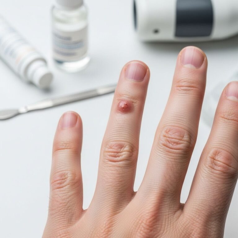
- Early nodular BCC on nose: Small, shiny pink papule with telangiectasia (Image 1).
- Advanced nodular BCC on cheek: Pearly nodule with central ulceration and rolled borders (Image 2).
- Pigmented nodular BCC on forehead: Dome-shaped lesion with blue-gray pigmentation (Image 3).
- Ulcerated nodular BCC on temple: Large crateriform ulcer with heaped-up edges (Image 4).
- Multiple nodular BCCs in fair-skinned patient: Cluster on sun-damaged back (Image 5).
These images highlight the variability; always consult a dermatologist for biopsy confirmation.
Dermoscopy of nodular BCC
Dermoscopy enhances diagnostic accuracy for nodular BCC, revealing specific vascular and structural patterns. Non-pigmented lesions show absence of pigment network plus at least one of: ulceration, arborising telangiectasias, blue-gray globules, leaf-like areas, or spoke-wheel areas.
Dermoscopic criteria table:
| Feature | Non-pigmented BCC | Pigmented BCC | Superficial BCC (for comparison) |
|---|---|---|---|
| Pigmentation | Lack of pigment network | Blue-gray ovoid nests, globules | Shiny white-red areas |
| Vessels | Arborising telangiectasias | Arborising vessels | Short fine telangiectasias |
| Surface changes | Ulceration, erosions | Leaf-like areas | Erosions, arborising |
Typical dermoscopic images include:
- Arborising vessels branching like tree roots (high specificity).
- Ulceration with white structureless areas.
- In pigmented variants: large blue-gray ovoid nests (D4 criterion).
Histopathology of nodular BCC
Histologically, nodular BCC shows lobules of basaloid cells extending from the epidermis into dermis. Key features:
- Peripheral palisading: orderly alignment of nuclei at tumour edges.
- Retraction artefact: clefts between tumour and stroma due to shrinkage.
- Mucinous stroma and cystic degeneration in larger nodules.
- No atypia in epidermis; tumour connected to basal layer.
Immunohistochemistry: Positive for BerEP4, bcl-2, CD10; p53 in aggressive cases. Nodulocystic variant has central necrosis.
Histological images depict:
- Low-power view: Nodular aggregates in dermis (Image H1).
- High-power: Palisading and retraction (Image H2).
Diagnosis of nodular BCC
Diagnosis combines clinical exam, dermoscopy, and biopsy. Suspicious lesions warrant punch or shave biopsy to confirm subtype and risk (low vs. high-risk based on size, site, depth). High-risk features: face, >2 cm, ill-defined borders, recurrence.
Differential diagnosis
Nodular BCC mimics:
- Benign: Sebaceous cyst, trichoepithelioma, basaloid follicular hamartoma.
- Malignant: Basaloid squamous cell carcinoma, Merkel cell carcinoma (use IHC).
- Other: Amelanotic melanoma, pyogenic granuloma.
Dermoscopy and biopsy differentiate reliably.
Treatment and management
Standard treatment is surgical excision with 4–5 mm margins for low-risk nodular BCC (cure rate >95%). Mohs micrographic surgery for high-risk sites (face, eyes). Alternatives: Curettage/electrodesiccation for small lesions, cryotherapy, topical imiquimod, or radiotherapy.
Follow-up: Every 6–12 months lifelong due to 5–10% recurrence and new BCC risk. Sun protection prevents recurrence.
Frequently Asked Questions
What does nodular BCC look like?
A shiny, pearly nodule with rolled edges and telangiectasias, often ulcerating centrally.
Is nodular BCC dangerous?
Locally invasive if untreated but rarely metastasises; early treatment curative.
How is nodular BCC diagnosed?
Clinical exam, dermoscopy, and biopsy.
Can nodular BCC be cured?
Yes, with surgery; >95% cure rate for primary lesions.
What are risk factors for nodular BCC?
UV exposure, fair skin, age >40, immunosuppression.
See more basal cell carcinoma images.
References
- Basal cell carcinoma – diagnosis — Skvara H et al. PMC. 2014-02-12. https://pmc.ncbi.nlm.nih.gov/articles/PMC3934050/
- Nodular Basal Cell Carcinoma: Signs, Risks & Treatment — GentleCure. 2023. https://www.gentlecure.com/nodular-basal-cell-carcinoma/
- Basal and Squamous Cell Skin Cancer Symptoms — American Cancer Society. 2025-01-15. https://www.cancer.org/cancer/types/basal-and-squamous-cell-skin-cancer/detection-diagnosis-staging/signs-and-symptoms.html
- Basal Cell Carcinoma Untreated for 2 Years — Dermatology Seattle. 2024. https://dermatologyseattle.com/basal-cell-carcinoma-untreated-for-2-years-what-you-need-to-know/
- Basal cell carcinoma – Symptoms & causes — Mayo Clinic. 2025-06-10. https://www.mayoclinic.org/diseases-conditions/basal-cell-carcinoma/symptoms-causes/syc-20354187
- Basal Cell Carcinoma — Cleveland Clinic. 2024-11-20. https://my.clevelandclinic.org/health/diseases/4581-basal-cell-carcinoma
Similar Articles


Read full bio of Sneha Tete