Normal Wound Healing: 4 Phases And Key Mechanisms
Comprehensive guide to the phases, processes, and factors influencing normal wound healing in human skin.

The process of normal wound healing is a complex, dynamic sequence of biological events that restores the integrity of the skin following injury. It involves coordinated cellular and molecular interactions progressing through distinct but overlapping phases: haemostasis, inflammation, repair (proliferation), and maturation (remodelling). This article details each phase, key mechanisms such as epithelialisation, fibroplasia, and capillary proliferation, and factors influencing healing outcomes.
Wound Assessment
Assessing a wound is crucial for predicting healing potential and guiding management. Wound severity depends on
surface area
anddepth
. Partial-thickness wounds, affecting only the epidermis and upper dermis, heal faster by recruiting keratinocytes from adjacent adnexal structures like hair follicles and sweat glands. In contrast, full-thickness wounds, extending through the entire dermis, rely solely on keratinocyte migration from wound edges, leading to slower closure and potential scarring.Clinically, wounds are classified by depth:
- Superficial: Epidermis only; heals rapidly without scarring.
- Partial thickness: Epidermis and partial dermis; regenerates with minimal fibrosis.
- Full thickness: Entire dermis and possibly subcutaneous tissue; heals by contraction and scarring.
Additional factors include contamination, location (e.g., lower extremities heal slower due to poor perfusion), and patient comorbidities like diabetes or malnutrition.
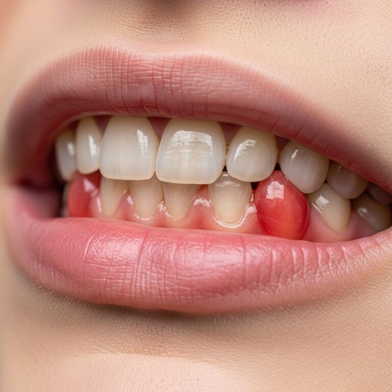
Haemostasis
Haemostasis is the immediate response to injury, lasting minutes to hours, aimed at stopping bleeding and forming a provisional matrix. Vascular constriction reduces blood flow, followed by platelet activation and aggregation. Platelets release clotting factors, forming a fibrin clot that seals the wound and provides a scaffold for cell migration.
This phase establishes the foundation for healing: the scab protects underlying tissues while platelets release growth factors like PDGF and TGF-β, recruiting inflammatory cells. Disruption, such as in haemophilia, prolongs bleeding and delays healing.
Inflammation
The inflammatory phase (days 1-6) clears debris and prevents infection. Vasodilation increases blood flow, delivering neutrophils and macrophages. Neutrophils phagocytose bacteria and dead tissue, while macrophages remove remaining debris and secrete cytokines (e.g., TNF-α, IL-1) and growth factors (e.g., VEGF, FGF) to stimulate repair.
Signs include redness, swelling, heat, and pain. Prolonged inflammation, due to infection or foreign bodies, releases excess proteases and reactive oxygen species, impairing healing. Resolution transitions to proliferation as macrophage numbers peak around day 3-4.
Repair
Repair, or proliferative phase (days 4-21), rebuilds tissue via epithelialisation, fibroplasia, and capillary proliferation (angiogenesis). Fibroblasts migrate into the wound, synthesizing collagen (types I and III) and extracellular matrix (ECM) components like fibronectin and proteoglycans, forming granulation tissue—a pink, vascular matrix.
Epithelialisation
Epithelialisation begins within hours: keratinocytes at wound edges lose adhesion (via desmosome dissolution), proliferate, and migrate over the provisional matrix under EGF, KGF, and TGF-α influence. A new basement membrane forms, restoring the epidermal barrier. Partial-thickness wounds re-epithelialise from adnexa; full-thickness from edges only.
Fibroplasia
Fibroblasts, activated by PDGF, produce disorganized ECM. Myofibroblasts, with contractile properties, enable wound contraction, reducing wound size by up to 80% in open wounds. Collagen cross-linking increases tensile strength.
Capillary Proliferation (Angiogenesis)
New capillaries sprout from wound edges, driven by VEGF and bFGF from macrophages and keratinocytes. This vascularizes granulation tissue, supplying oxygen and nutrients. Hypoxia-inducible factors regulate this process.
By week’s end, the wound surface closes, though underlying strength is only 20-30% of normal skin.
Maturation (Remodelling)
Maturation (weeks to years) refines the scar. Collagen reorganizes from type III to type I, increasing tensile strength to 80% of normal by 1 year. Matrix metalloproteinases (MMPs) from fibroblasts balance synthesis and degradation. Vessels regress via apoptosis, reducing hyperaemia. Myofibroblasts apoptose, halting contraction.
Large wounds may take years; scars remain avascular and hypopigmented. Fetal wounds regenerate without scarring due to minimal inflammation and hyaluronic acid-rich ECM.
Cells Involved in Wound Healing
| Cell Type | Phase | Key Functions |
|---|---|---|
| Platelets | Haemostasis | Clot formation, growth factor release (PDGF, TGF-β) |
| Neutrophils | Inflammation | Phagocytosis of bacteria/debris |
| Macrophages | Inflammation/Proliferation | Debridement, cytokine/growth factor secretion |
| Fibroblasts/Myofibroblasts | Proliferation/Maturation | ECM synthesis, contraction, remodelling |
| Keratinocytes | Proliferation | Migration, proliferation, barrier reformation |
| Endothelial cells | Proliferation | Angiogenesis |
This table summarizes primary cells; interactions via integrins and adhesion molecules orchestrate healing.
Mediators of Wound Healing
Growth factors and cytokines regulate healing:
- PDGF: Fibroblast recruitment/proliferation.
- TGF-β: ECM production, angiogenesis inhibition in maturation.
- VEGF: Angiogenesis.
- EGF/KGF: Epithelial proliferation/migration.
- Cytokines (IL-1, TNF-α): Inflammation amplification.
ECM components (collagen, fibronectin) provide structural support and signalling.
Frequently Asked Questions (FAQs)
What are the four phases of normal wound healing?
Haemostasis, inflammation, proliferation (repair), and maturation (remodelling).
How long does each phase last?
Haemostasis: minutes-hours; Inflammation: 1-6 days; Proliferation: 4-21 days; Maturation: weeks-years.
Why do some wounds scar?
Full-thickness wounds heal by contraction and fibrosis, lacking adnexal regeneration sources.
What delays healing?
Infection, poor nutrition, diabetes, smoking, or excessive inflammation.
Can wounds heal without scarring?
Fetal wounds do via regeneration; adult superficial wounds minimize it.
Clinical Implications
Understanding normal healing optimizes care: clean wounds promote inflammation resolution; moist environments enhance epithelialisation; offloading reduces shear. Chronic wounds fail phase progression, requiring targeted therapies. Optimal healing restores function with minimal deformity.
References
- Wound healing — Healthify.nz. 2023. https://healthify.nz/health-a-z/w/wound-healing
- Principles of Wound Healing – Mechanisms of Vascular Disease — NCBI Bookshelf (StatPearls). 2023-11-03. https://www.ncbi.nlm.nih.gov/books/NBK534261/
- The Four Stages of Wound Healing — Wound Evolution. 2023. https://www.woundevolution.com/blog/the-four-stages-of-wound-healing/
- Normal wound healing – DermNet — DermNet NZ. 2023. https://dermnetnz.org/cme/wound-healing/normal-wound-healing
- Wounds – DermNet — DermNet NZ. 2023. https://dermnetnz.org/topics/wounds
Similar Articles


Read full bio of Sneha Tete