Ocular Melanoma: Understanding Eye Cancer
Complete guide to recognizing, understanding, and managing eye melanoma

Ocular melanoma represents a serious but uncommon form of cancer that develops within the eye itself. Unlike skin melanoma, which is widely recognized and frequently discussed in health contexts, eye melanoma often develops silently without producing noticeable symptoms in its early stages. This characteristic makes it particularly challenging to detect, which is why understanding the disease and recognizing warning signs becomes essential for protecting your vision.
What Defines Ocular Melanoma
Ocular melanoma is a malignant tumor arising from melanocytes, the cells responsible for producing melanin, the pigment that provides color to our eyes, hair, and skin. This form of cancer occurs in approximately 5 out of every million adults, making it an exceptionally rare diagnosis. Despite its rarity, ocular melanoma represents the second most common type of melanoma overall, accounting for roughly 5% of all melanoma cases.
The disease is fundamentally different from skin melanoma. While cutaneous melanoma, the far more common skin-based variant, is strongly associated with sun exposure and UV radiation, ocular melanoma does not share this clear causal link with ultraviolet light. This distinction is crucial for understanding prevention strategies and risk assessment. The cancer develops within the eye’s delicate structures, making it both difficult to detect and challenging to treat without potentially affecting vision.
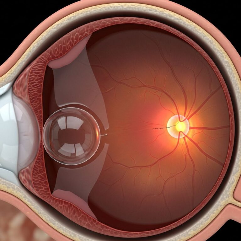
Anatomical Locations Within the Eye
Ocular melanoma can originate in multiple locations throughout the eye, though certain areas are affected more frequently than others. Understanding where these tumors develop helps explain why symptoms vary considerably between patients.
The Iris and Anterior Chamber
The iris, the colored portion of the eye that surrounds the pupil, represents one potential site for melanoma development. Melanomas originating in the iris are among the more visible types, as they can appear as growing dark spots on the colored part of the eye. Because the iris is located at the front of the eye, tumors here may be detected earlier than those in other regions, potentially improving outcomes. Iris melanomas represent approximately 10-15% of all ocular melanomas, making them relatively uncommon compared to other eye locations.
The Ciliary Body
Located behind the iris, the ciliary body serves multiple critical functions for eye health and vision. This tissue layer contains muscle fibers responsible for changing the lens shape to focus on objects at varying distances, and it also produces aqueous humor, the transparent fluid that maintains eye pressure and nourishes the cornea and lens. When melanoma develops in the ciliary body, it may disrupt these important functions, potentially leading to blurred vision or sudden changes in eyeglass prescription requirements. Ciliary body melanomas account for a significant portion of ocular melanomas and often reach a substantial size before detection.
The Choroid Layer
The choroid, positioned between the retina and the sclera (white outer layer), represents the most common site for ocular melanoma development. This vascular tissue layer is rich in blood vessels and pigmented cells that nourish the retina, the light-sensitive tissue essential for vision. Choroidal melanomas represent approximately 80-85% of all ocular melanomas. Because the choroid lies behind the retina and cannot be seen by looking in a mirror, tumors in this location often remain undetected until they grow large enough to affect vision or are discovered during routine eye examinations.
Recognizing Symptoms and Warning Signs
Many individuals with early-stage ocular melanoma experience no symptoms whatsoever, making routine eye examinations critical for early detection. However, as tumors progress, various vision-related symptoms may emerge.
Common Visual Symptoms
- Floaters and Light Flashes: Patients often report seeing specks of dust or floaters in their visual field, along with occasional flashes of light. These phenomena result from the tumor’s effects on the retina or vitreous humor.
- Blurred or Impaired Vision: Many individuals experience blurriness or unusually poor vision, typically affecting one eye. This symptom may develop gradually or appear suddenly.
- Peripheral Vision Loss: Loss of side vision occurs when tumors damage the peripheral retina or affect the optic nerve.
- Dark Spots on the Iris: A growing dark spot visible on the colored part of the eye may indicate iris melanoma.
- Pupil Changes: Alterations in pupil shape or size can result from ciliary body involvement or pressure from expanding tumors.
- Eye Bulging or Displacement: Larger tumors may cause the eye to protrude or shift position within the socket.
- Distorted Vision: Some patients report that straight lines appear wavy or that portions of visual grids seem missing, particularly when choroidal melanomas affect the macula.
Additional Symptoms
Beyond vision changes, some patients experience eye irritation, pain, redness, or a sensation of something foreign in the eye. Increased pressure within the eye may also occur. If a choroidal melanoma causes retinal detachment, vision problems can escalate rapidly. These diverse symptoms underscore why comprehensive eye examinations form the cornerstone of early detection.
Understanding Risk Factors
While the exact cause of ocular melanoma remains incompletely understood, researchers have identified several factors that increase risk. It is crucial to note that possessing risk factors does not guarantee disease development—many individuals with multiple risk factors never develop melanoma, while some without recognized risk factors do.
Demographic and Genetic Factors
Caucasian individuals face significantly higher risk compared to other populations. Age represents another important factor, with ocular melanoma occurring more frequently in older adults. Individuals with light-colored eyes, including blue, green, or light brown eyes, carry elevated risk. Additionally, people with fair skin, fair or red hair, and blue eyes—the same phenotypic characteristics associated with increased skin melanoma risk—show higher susceptibility.
Pre-existing Eye and Skin Conditions
Certain inherited conditions substantially increase ocular melanoma risk. Dysplastic nevus syndrome, a condition characterized by unusual moles on the skin, raises melanoma risk in both skin and eyes. Similarly, ocular melanocytosis, involving unusual skin pigmentation on the eyelids and surrounding tissues coupled with increased pigmentation on the uvea, significantly elevates risk.
Environmental Exposures
Unlike skin melanoma, ocular melanoma shows no conclusive evidence linking to sun exposure or ultraviolet radiation. However, some research suggests that frequent UV light exposure may modestly increase risk, though this relationship remains unproven and debated among researchers.
How Ocular Melanoma Develops
At the cellular level, ocular melanoma develops when errors or mutations in the DNA of melanocytes cause these cells to multiply uncontrollably. These genetic abnormalities lead cells to ignore normal growth-limiting signals, resulting in progressive tumor expansion. Scientists believe that a combination of genetic predisposition and environmental factors contributes to these DNA errors, though the specific mechanisms remain incompletely elucidated.
The aggressive nature of ocular melanoma relates partly to its vascularity. Because the uvea contains rich blood supplies, melanomas in these locations have abundant access to nutrients and oxygen, potentially facilitating rapid growth. Additionally, the eye’s location and the tumor’s access to blood vessels create conditions favorable for metastasis, where cancer cells spread to distant organs.
The Serious Risk of Metastasis
One of the most concerning aspects of ocular melanoma involves its potential to spread beyond the eye. In approximately half of all ocular melanoma cases, the cancer metastasizes to distant organs, most commonly the liver. This metastatic spread often proves fatal, making it the primary cause of death in ocular melanoma patients. The aggressive nature of the disease and its propensity for late metastatic presentation underscore the importance of early detection and appropriate treatment.
Diagnostic Approaches
Because early-stage ocular melanoma typically produces no symptoms, diagnosis often occurs incidentally during routine eye examinations. Eye care professionals use specialized equipment and techniques to identify suspicious lesions. Detailed imaging studies, including ultrasound and MRI, help determine tumor size, location, and extent. These diagnostic tools are essential for staging the cancer and planning appropriate treatment strategies.
Treatment Considerations
Treatment approaches for ocular melanoma vary based on tumor size, location, and individual patient factors. Options may include radiation therapy, surgical removal of the tumor, or in advanced cases, removal of the entire eye (enucleation). The goal of treatment involves eliminating cancer while preserving vision and eye function whenever possible. Discussions between patients and their ophthalmologists regarding treatment benefits and risks are essential for making informed decisions.
Importance of Regular Eye Examinations
Given that ocular melanoma often develops without symptoms, regular comprehensive eye examinations represent the most effective strategy for early detection. Adults should have their eyes examined by qualified eye care professionals at intervals recommended based on age, health status, and risk factors. Individuals with known risk factors or a family history of ocular melanoma may benefit from more frequent screening.
Living with Ocular Melanoma
A diagnosis of ocular melanoma requires adjustment to living with a serious health condition. Patients benefit from working closely with experienced ophthalmologists and oncologists who specialize in intraocular malignancies. Support groups and patient advocacy organizations provide valuable resources for navigating treatment decisions, managing side effects, and connecting with others facing similar challenges. Regular follow-up examinations and monitoring remain essential even after initial treatment to detect any recurrence or metastatic spread.
Key Takeaways
- Ocular melanoma is a rare form of eye cancer arising from melanin-producing cells within the eye
- The disease often develops without initial symptoms, making routine eye exams crucial for detection
- Multiple anatomical locations within the eye can develop melanoma, with the choroid being most common
- Risk factors include Caucasian ancestry, light eye color, age, and certain inherited skin conditions
- Unlike skin melanoma, ocular melanoma shows no clear link to sun exposure
- Metastasis to the liver and other organs occurs in approximately half of cases
- Early detection through regular eye examinations significantly impacts treatment outcomes
- Treatment options vary based on tumor characteristics and may include radiation or surgery
Frequently Asked Questions
Is ocular melanoma preventable?
While certain risk factors like genetic predisposition cannot be changed, there is no proven prevention strategy for ocular melanoma. Unlike skin melanoma, sun protection does not conclusively reduce risk. The best approach involves regular eye examinations for early detection, particularly for individuals with known risk factors.
Can ocular melanoma be detected without symptoms?
Yes, many ocular melanomas are detected during routine eye examinations before the patient experiences any symptoms. This is why regular comprehensive eye exams are so important, especially for those with risk factors.
How is ocular melanoma different from skin melanoma?
While both arise from melanocytes, ocular melanoma is not related to sun exposure as skin melanoma is. The two conditions represent distinct diseases with different risk factors and management approaches. Ocular melanoma is also much rarer than cutaneous melanoma.
What is the prognosis for ocular melanoma?
Prognosis depends on tumor size, location, and whether metastasis has occurred. Early detection generally offers better outcomes. However, the risk of eventual metastasis, particularly to the liver, remains significant even after successful local treatment.
Will I lose my vision if diagnosed with ocular melanoma?
Vision loss depends on tumor location and size. Some patients retain good vision, while others experience vision impairment. Treatment approaches aim to eliminate cancer while preserving vision when possible, though this is not always achievable.
References
- Eye melanoma – Symptoms and causes — Mayo Clinic. 2024. https://www.mayoclinic.org/diseases-conditions/eye-melanoma/symptoms-causes/syc-20372371
- Ocular (Eye) Melanoma – Symptoms and Causes — Penn Medicine. 2024. https://www.pennmedicine.org/conditions/ocular-melanoma
- Ocular Melanoma – Symptoms, Causes, Treatment — National Organization for Rare Disorders (NORD). 2024. https://rarediseases.org/rare-diseases/ocular-melanoma/
- Ocular Melanoma Foundation – Disease — Ocular Melanoma Foundation. 2024. https://ocularmelanoma.org/disease
- Ocular Melanoma – Melanoma — Moffitt Cancer Center. 2024. https://www.moffitt.org/cancers/melanoma/diagnosis/types/ocular-melanoma/
Similar Articles


Read full bio of medha deb