Onychomatricoma Explained: Key Causes, Diagnosis, & Treatments
Rare benign nail matrix tumour causing nail thickening, yellow bands, and splinter haemorrhages in adults.

Onychomatricoma is a rare, benign fibroepithelial tumour originating from the nail matrix, first described by Baran and Kint in 1992. This condition primarily affects the fingernails, leading to distinctive changes in nail appearance and structure that can mimic more common nail disorders.
Who gets onychomatricoma?
Onychomatricoma typically presents in middle-aged Caucasian women, with peak incidence around the fifth decade of life. It most frequently involves the thumb or index finger of the dominant hand, though cases on toes, including the great toenail, have been reported. The tumour is exceptionally rare in children and shows no strong predisposition in other ethnic groups.
- Demographic profile: Predominantly women over 40 years old.
- Site preference: First three fingernails of dominant hand (thumb most common).
- Rarity: Fewer than 80 cases documented worldwide since initial description.
Causes
The precise aetiology of onychomatricoma remains unclear, but associations with prior nail trauma and coexisting onychomycosis (fungal nail infection) have been noted in several cases. Recent genetic studies suggest possible chromosomal alterations, such as deletions, in affected patients, indicating a potential molecular basis. However, no consistent causative factor has been identified, and the tumour is considered sporadic.
Onychomycosis complicating onychomatricoma can exacerbate symptoms and lead to misdiagnosis, as fungal elements may colonise the abnormal nail structure.
Clinical features
The classic clinical tetrad of onychomatricoma includes:
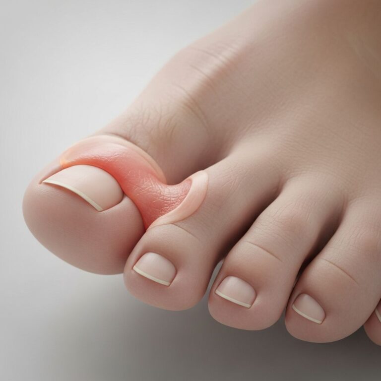
- Thickening of the nail plate: Localized or diffuse hypertrophy, often 2-3 times normal thickness.
- Transverse or longitudinal overcurvature: Spoon-like or funnel-shaped nail deformity.
- Xanthonychia: Yellow longitudinal band(s) of variable width, sometimes with red or brown pigmentation.
- Multiple splinter haemorrhages: Longitudinal red streaks, especially prominent distally.
Additional features may include swelling of the proximal nail fold, nail dystrophy, dorsal pterygium (scar-like fusion of proximal nail fold to nail plate), and honeycomb-like cavities visible at the distal nail margin upon avulsion. The nail plate often exhibits ridging, splitting, or pitting, and the lesion is typically painless unless secondarily infected.
| Feature | Description | Frequency |
|---|---|---|
| Nail thickening | Diffuse or localised hypertrophy | Nearly universal |
| Overcurvature | Transverse/longitudinal | Common |
| Xanthonychia | Yellow band | Highly characteristic |
| Splinter haemorrhages | Multiple longitudinal | Classic sign |
Complications
Onychomatricoma is benign but can lead to secondary onychomycosis due to altered nail architecture trapping debris and fungi. Misdiagnosis as fungal infection delays appropriate treatment, potentially causing prolonged dystrophy. Rarely, nail matrix damage post-excision may result in permanent dystrophy, though recurrence is uncommon with complete removal.
- Fungal superinfection (onychomycosis).
- Pain from trauma to thickened nail.
- Cosmetic deformity and functional impairment.
Diagnosis
Diagnosis combines clinical suspicion with dermoscopy, imaging, and confirmatory histopathology. After nail avulsion, the tumour appears as a villous matrix lesion with finger-like projections penetrating the nail plate, creating characteristic cavities.
Dermoscopy
Reveals perforations in distal nail plate, white longitudinal grooves, parallel lesion edges, dark dots, nail pitting, and haemorrhagic striae.
Imaging
- Ultrasound: Hypoechoic tumour in matrix with hyperechoic finger-like projections and low vascularity.
- MRI: Y-shaped proximal nail plate thickening and transverse perforations.
- X-ray: No bone involvement.
Histopathology
Definitive diagnosis shows a biphasic fibroepithelial tumour:
- Proximal zone: Deep epithelial invaginations beneath proximal nail fold filled with ungual protrusions.
- Distal zone (lunula): Epithelial digitations perforating nail plate, with fibrous cores and thin epithelium.
Immunohistochemistry may highlight matrix-specific keratin expression.
Differential diagnoses
| Condition | Key Distinguishing Features |
|---|---|
| Onychomycosis | Mycological confirmation; no villous projections on avulsion. |
| Longitudinal melanonychia | Pigment-focused; dermoscopy shows melanocytic pattern. |
| Fibrokeratoma / Periungual fibroma | Localized fibrous nodule; no matrix involvement. |
| Squamous cell carcinoma / Bowen’s disease | Destructive growth, pain; biopsy differentiates. |
| Osteochondroma / Verruca | Bony/radiological changes or HPV features. |
Treatment
Surgical excision is the standard treatment, requiring complete removal of the tumour including adjacent normal nail matrix to prevent recurrence. Procedure:
- Local anaesthesia and nail plate avulsion.
- Visualization and excision of villous projections from matrix.
- Margin inclusion of healthy matrix tissue.
Minimally invasive techniques have been reported successfully. Post-excision, normal nail regrowth occurs within months if matrix preserved adequately. Recurrence is rare (one case in mean 20-month follow-up).
Outcome
Prognosis is excellent following complete excision, with low recurrence risk and good cosmetic/functional recovery. Persistent dystrophy may occur if matrix extensively involved. Early diagnosis prevents complications like superinfection.
Frequently Asked Questions (FAQs)
Is onychomatricoma painful?
Typically painless, but may become tender if traumatised or infected.
Can onychomatricoma affect toenails?
Yes, though less common than fingernails; great toenail reported.
Does onychomatricoma turn cancerous?
No, it is benign with no malignant potential.
How is onychomatricoma diagnosed without surgery?
Dermoscopy and ultrasound strongly suggest it; histopathology confirms post-excision.
What if misdiagnosed as fungus?
Antifungals fail; persistent symptoms prompt re-evaluation.
References
- Onychomatricoma — DermNet NZ. 2023. https://dermnetnz.org/topics/onychomatricoma
- Onychomatricoma — Orphanet. 2023-10-01. https://www.orpha.net/en/disease/detail/300512
- Onychomatricoma: A Rare Tumor of Nail Matrix — PMC/NIH (Ann Dermatol). 2016-04-01. https://pmc.ncbi.nlm.nih.gov/articles/PMC4828389/
- Clinical Presentation and Diagnostic Features of Onychomatricoma: A Case Report — EMJ Reviews (Dermatology). 2023. https://www.emjreviews.com/dermatology/abstract/clinical-presentation-and-diagnostic-features-of-onychomatricoma-a-case-report-j030123/
- Onychomatricoma Presenting as a Dystrophic Right Great Toenail — PubMed (Skin Appendage Disord). 2020-06-01. https://pubmed.ncbi.nlm.nih.gov/32499986/
- Onychomatricoma – Wikipedia — Wikipedia. 2024. https://en.wikipedia.org/wiki/Onychomatricoma
Similar Articles


Read full bio of Sneha Tete