Open-Angle Glaucoma: Symptoms, Diagnosis, Treatment Guide
Understand open-angle glaucoma: the silent thief of sight, its causes, symptoms, diagnosis, and life-saving treatments.

Open-angle glaucoma (OAG) is the most common form of glaucoma, a chronic and progressive optic neuropathy that leads to irreversible vision loss if untreated. Characterized by an open anterior chamber angle, it damages the optic nerve primarily due to elevated intraocular pressure (IOP), though normal-tension cases exist. Often dubbed the ‘silent thief of sight,’ it advances asymptomatically until advanced stages, making regular eye exams essential for early detection and management.
What Is Open-Angle Glaucoma?
Open-angle glaucoma occurs when the trabecular meshwork, the eye’s drainage system, becomes obstructed, impeding aqueous humor outflow despite an open angle between the iris and cornea. This leads to gradual IOP elevation above 21 mm Hg in most cases, compressing the optic nerve and causing retinal ganglion cell death. Primary open-angle glaucoma (POAG) lacks secondary causes, while secondary forms stem from trauma, inflammation, or pigmentation. The condition is bilateral but often asymmetric, progressing from peripheral to central vision loss.
Unlike acute angle-closure glaucoma, OAG lacks sudden symptoms, with damage accumulating over years. Central vision remains intact until late stages, when blindness can ensue. IOP is the primary modifiable risk factor, but fluctuations and levels below 22 mm Hg occur in 40-50% of cases, highlighting multifactorial etiology including vascular and genetic components.
Symptoms of Open-Angle Glaucoma
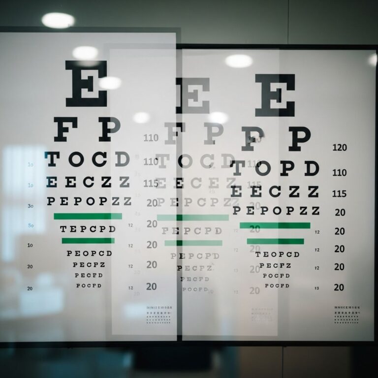
Early open-angle glaucoma is asymptomatic, evading detection without screening. As it advances, patients notice gradual peripheral vision loss, described as tunnel vision. Advanced symptoms include blurred vision, halos around lights, eye pain, and headaches from IOP spikes. Disc hemorrhages or nerve fiber layer defects signal active progression. Visual field testing reveals arcuate scotomas corresponding to optic disc rim thinning.
- Gradual loss of side (peripheral) vision
- Tunnel vision in moderate to advanced stages
- Blurry vision or halos, especially at night
- Mild eye pain or headaches (less common)
- No symptoms until 40-50% nerve fibers are lost
Risk of missing early disease is high; only advanced cases prompt medical attention. Routine exams detect pre-symptomatic changes via tonometry and optic nerve assessment.
Risk Factors for Open-Angle Glaucoma
Multiple factors elevate OAG risk, with IOP as the most significant modifiable element. Population studies confirm higher prevalence with IOP >21 mm Hg; Ocular Hypertension Treatment Study (OHTS) data show progression risk doubling per 1 mm Hg increase.
| Risk Factor | Description | Relative Risk |
|---|---|---|
| Elevated IOP | >21 mm Hg; fluctuations worsen progression | Primary modifiable factor |
| Age >60 | Prevalence rises exponentially after 40 | 10x higher in >80 years |
| Family History | First-degree relative increases risk 4-9x | Genetic predisposition |
| African American Race | Earlier onset, higher prevalence | 4-5x vs. Caucasians |
| Thin Corneas | Underestimates true IOP | 2x progression risk |
| Myopia/High Eye Pressure | Large cups, vascular issues | Elevated |
Other contributors: diabetes, hypertension, steroid use, eye trauma. Normal-tension glaucoma affects those with IOP <21 mm Hg, linked to vascular dysregulation.
How Is Open-Angle Glaucoma Diagnosed?
OAG diagnosis is multifaceted, excluding angle-closure or secondary causes. It requires open anterior chamber angle, optic neuropathy signs, and visual field defects. No single test suffices; glaucoma suspects need monitoring.
Gonioscopy: Gold standard confirms open angle (20-45°). Differentiates from narrow-angle disease.
Tonometry: Measures IOP; diurnal variation testing advised. Pachymetry corrects for corneal thickness.
Ophthalmoscopy/Optic Nerve Imaging: Enlarged cup-to-disc ratio (>0.6), asymmetry (>0.2), notching, hemorrhages, peripapillary atrophy. OCT quantifies retinal nerve fiber layer thinning.
Visual Field Testing (Perimetry): Standard automated perimetry detects scotomas. Early: nasal steps; advanced: arcuate defects near fixation.
- Mild: Optic disc/RNFL damage, normal fields
- Moderate: Field loss >5° from fixation in one hemifield
- Severe: Field loss in both hemifields or near fixation
Staging guides therapy; progression monitoring every 3-6 months.
Treatment for Open-Angle Glaucoma
Treatment targets IOP reduction by 20-50%, halting progression. Early Manifest Glaucoma Trial showed 10% risk reduction per mm Hg lowered. Options: medications, laser, surgery.
Medications (First-Line):
- Prostaglandin analogs (latanoprost): 25-35% IOP drop, nightly
- Beta-blockers (timolol): 20-25% reduction, twice daily
- Alpha agonists, carbonic anhydrase inhibitors as adjuncts
Laser Trabeculoplasty: Selective laser (SLT) boosts trabecular outflow; repeatable, minimal downtime.
Surgery: Trabeculectomy, tube shunts, MIGS (minimally invasive) for advanced/refractory cases. Enhances drainage via new pathways.
Tailor to stage: mild (meds), moderate (laser + meds), severe (surgery). Monitor progression; noncompliance common barrier.
Prevention and Prognosis
No cure exists, but early intervention preserves vision. Annual exams post-40, especially high-risk groups. Lifestyle: cardio exercise lowers IOP, avoid steroids. Prognosis excellent with compliance; untreated, 53% progress in 6 years.
Interprofessional care optimizes outcomes: ophthalmologists, optometrists, nurses educate on adherence.
Frequently Asked Questions (FAQs)
Can open-angle glaucoma be cured?
No, it’s irreversible, but treatments prevent further loss. Early detection is key.
At what age should screening begin?
Age 40 for general; earlier (30s) for high-risk (family history, African American).
Does high IOP always mean glaucoma?
No, ocular hypertension may not progress; monitor suspects.
Is laser treatment permanent?
SLT effects last 1-5 years; repeatable unlike ALT.
Can lifestyle changes help?
Exercise, diet control systemic risks; no substitute for meds.
References
- Open Angle Glaucoma – StatPearls — NCBI Bookshelf/NCBI Staff. 2023-08-08. https://www.ncbi.nlm.nih.gov/books/NBK441887/
- Primary Open-Angle Glaucoma — EyeWiki/American Academy of Ophthalmology. 2024-05-15. https://eyewiki.org/Primary_Open-Angle_Glaucoma
- Glaucoma Risks — Glaucoma Research Foundation. 2023-11-20. https://glaucoma.org/understanding-glaucoma/risks
Similar Articles


Read full bio of medha deb